| Video: Colorectal Cancer Overview |
Overview
Overview Colon cancer is the second most commonly diagnosed cancer. 1/12 people will develop bowel cancer before the age of 85. However, there are Survival rates are increasing. Early bowel cancer is cured by surgery alone (screening is important!!). If untreated, or diagnosed when distance metastases are present, >98% pf patients die in <5 years.
| Definition Polyps: protuberance into the lumen of normally flat colonic mucosa Colorectal Cancer: Cancer of the colon and/or rectum usually at an advanced age. It is an adenoma and the primary site of metastasis is the liver. Familial Adenomatous Polyposis: autosomal dominant inheritance (mutation of the adenomatous polyposis coli gene), characterised by hundred to thousands of colorectal adenomas usually by age 20. Lynch Syndrome: autosomal dominant inheritance (mutation of the mismatch repair gene) predisposing one to develop cancer at a younger age. |
Colon anatomy and Physiology
The colon is divided into:
- Caecum (appendix off this)
- Ascending colon
- Transverse colon
- Descending colon
- Sigmoid colon
- Rectum
The ascending and descending colon are retroperitoneal, these are immobile. The transverse is mobile and lies within the peritoneal cavity. The transverse colon is supported by the greater omentum superiorly and attaches to the posterior abdominal wall (in front of the retroperitoneal cavity) by the transverse mesocolon.
The transverse colon is attached to the greater curvature of the stomach and first part of the duodenum via the greater omentum. The sigmoid colon is also attached
Main features of large intestine structure:
- Complete layer of circular smooth muscle throughout, but incomplete bands of longitudinal muscle (taeniae coli) in colon (stops at the rectum)
- Fatty appendages along taeniae (appendices epiploicae)
- Folded internal mucosal appearances (haustrations)
- ‘Segmented’ external appearances (sacculations)
Blood supply - from superior and inferior mesenteric artery
- Superior mesenteric artery
- Ileocolic artery - last terminal ilealloop, caecum
- Right colic artery - ascending colon
- Middle colic artery - transverse colon up to the splenic flexure
- Inferior mesenteric artery
- Left colic - splenic flexure and descending colon.
- Sigmoid artery - sigmoid colon
- Superior rectal artery - rectum and upper anal canal
Autonomic nerve supply
- Sympathetic, mainly from greater splanchnic nerves via SMA and IMA plexuses.
- Parasympathetic, from vagus via SMA and IMA plexus from caecum to splenic flexure and from pelvic parasympathetics (S2, 3, 4) via hypogastric plexuses and retroperitoneal nerves from splenic flexure to upper anal canal
| Watch The Colon Anatomy and Colon Cancer |
Risk Factors
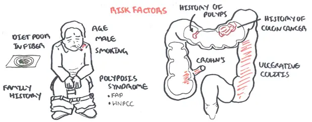
Risk Factor Family and personal history are strong risk factors. Polyposis syndrome including FAP has a 100% chance of developing colon cancer.
Signs and Symptoms
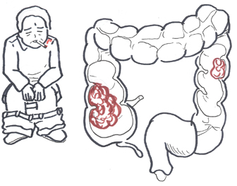
Signs and symptoms vary depending on the location of the tumour. 20% of people present at emergency with obstruction of large bowel or perforation
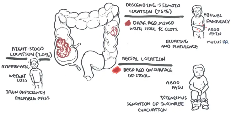
For details look at table below
| CLINICAL PRESENTATION | |||
| Right Colon | Left and Sigmoid Colon | Rectum | |
| Frequency | 20% | 75% | 5% |
| Pathology | Exophytic lesions wit occult bleeding | Annular, invasive lesions | Ulcerating |
| Symptoms | Weight los, weaknes, rarely obstruction | Constipation, change in bowel habits, abdominal pain, rectal bleeding | Obstruction, tenesumus, rectal bleeding |
| Signs | Fe+ deficiency anaemia, QLR mass (10%) | Palpable mass on Digital Rectal Examination | |
| Remember Elderly persons who present with iron-deficiency anemia should be investigated for colon cancer |
| Remember Occult blood in the stool of a person older than 40 years should be considered colon cancer until proven otherwise. To rule out colon cancer, perform a colonoscopy. |
Clinical Examination
- Anaemia
- Palpable mass on abdominal examination
- Palpable nodular liver (metastasis)
- Pigmentation (Peutz-Jeghers syndrome)
- Discrete black brown lesion on lips
Differential Diagnosis
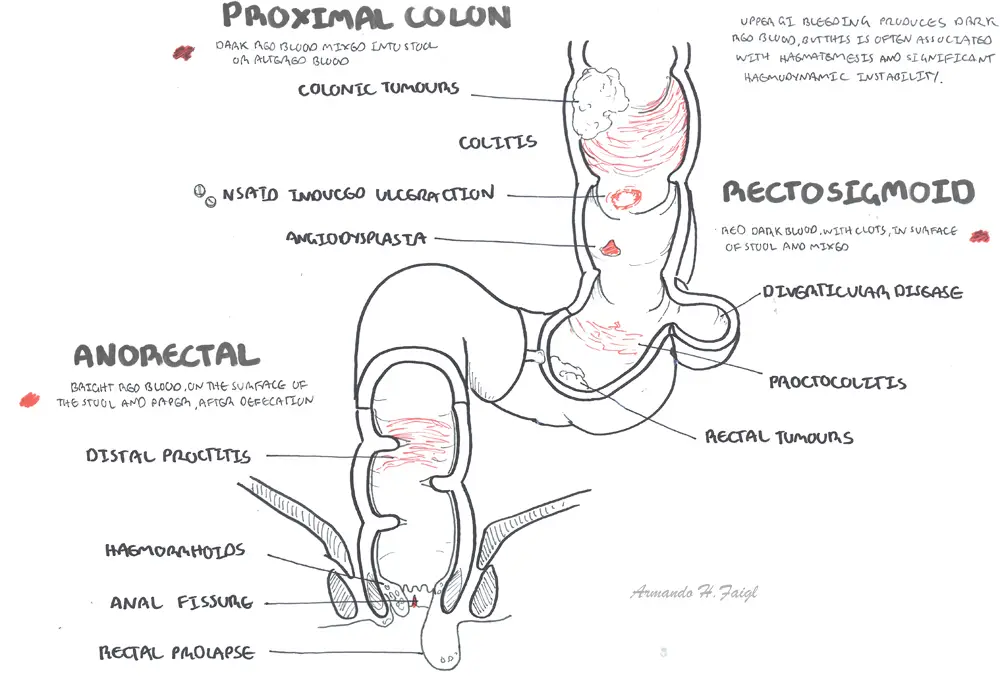
Differential Diagnosis of per-rectal bleeding
For more information: Clinical Presentation of Per-rectal bleeding
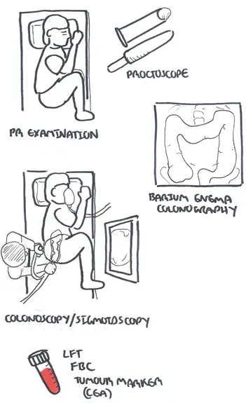
Investigation
- Rectal examination
- FBC
- Faecal occult blood testing (not done if symptomatic)
- Tumour markers
- CT imaging
- Colonoscopy
Investigation
| Remember Screening for colon cancer is available: FOBT. Done yearly after 50yo (Australia). |
Diagnosis Colon cancer is usually diagnosed with colonoscopy and biopsy (takes 1-2 days for pathology results)
Pathology
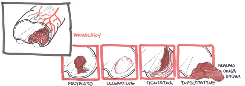
Pathology Colorectal cancer are primarily adenocarcinoma. Coloncancer can be polyploid, ulcerative, stenosing or infiltrative.
| Remember Blumer’s shelf: A firm lump felt in the perirectal pouch on rectal examination. It is a rare physical finding in patients with metastatic adenocarcinoma from the GIT usually the stomach. |
Pathophysiology
Staging and Management

Staging CT and PET scan are used for staging colon cancer. The staging system used is TNM which look at the primary tumour, regional lymph node involvement and metastasis. Duke's staging looks at the 5 year survival rate and this is based on the layers/sites the tumours have invaded.
Duke's staging (5 year survival rate)
- Stage A - Tumour is confined to bowel wall only (75-90%)
- Stage B - Tumour is through bowel wall (55-70%)
- Stage C - Tumour has spread through regional lymph nodes and has a 30% 5 year survival rate
- Stage D is where the tumour has metastasized has a 5% 5 year survival rate.
Management
- Surgical Resection
- +/- Neoadjuvant Chemotherapy
- Chemotherapy
- Radiotherapy
- Palliative care
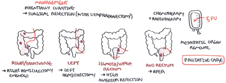
Management Surgery can be curative if done early with lymphadenectomy. Surgical removal of parts of the colon will depend on site of primary tumour(s). Chemoradiotherapy is also used, chemo agent primarily 5-flurouracil. Finally palliative care if surgery is not an option.
Complications and Prognosis
Complications
- Chemotherapy side effects
- Bone marrow suppression
- Nausea, diarrhoea, abdominal pain
- Alopecia
- Hand-foot Syndrome
| Hand-Foot Syndrome is a side effect for 5-fluorouracil chemotherapy characterised by a tender, symmetrical erythema of the palms and sole of foot. |
- Oxaliplatin side effects
- Neuropathy
- Hepatotoxicity
- Pulmonary fibrosis
- Surgical complications
- Haemorrhage
- Erectile dysfunction and/or bladder dysfunction (after rectal excision)
Prognosis (Based on Dukes) Criteria 5-year survival rates for colorectal cancer are:
- Stage I - 90%
- Stage II - 75%
- Stage III - 44% to 83%
- Stage IV - 8%
Prevention
Screening
- Genetic testing for those with hereditary conditions
- Faecal Occult blood test
- Colonoscopy?
Family Adenomatous Polyposis
Overview
- Autosomal dominant
- APC gene on chromosome 5q21
- Penetrance 100%
- Characterized by 100's of colonic polyps
- Mean age 39
Other manifestations
- Periemullary polyps
- Congenital hypertrophy of retinal pigment epithelium
- Osteomas skill and mandible
- Dental abnormalities
Hereditary Non-Poyposis colorectal Cancer
Overview
- Also known as Lynch Syndrome
- Autosomal dominant
- 80% lifetime risk of developing colorectal cancer
- increased risk uterine, gastric, urinary tract, small bowel and brain cancer
- Defect in DNA mismatch repair cause microsatellite instability
Peutz-Jeghers Syndrome
Overview
- Characterized by the development of noncancerous growths called hamartomatous polyps in the gastrointestinal tract (particularly the stomach and intestines)
- Greatly increased risk of developing certain types of cancer
- Increases risk of small-bowel and pancreatic cancer, colorectal cancer and sex-cord tumours with annular tubules of the ovary
- Other clinical features often found in children and young adults: Perioral pigmentation, pigmentations of fingers