Overview
Bowel obstruction is a blockage of intestinal contents, either mechanical (e.g., adhesions, hernias, malignancy) or functional (e.g., ileus), leading to impaired transit, proximal dilation, and downstream complications including ischaemia and perforation if untreated. Key features of bowel obstruction include colicky abdominal pain, distension, nausea with bilious or faeculent vomiting, obstipation, initially high-pitched then absent bowel sounds, and signs of dehydration or hypovolaemia.
Common in postoperative patients, older adults, and individuals with prior abdominal surgeries. Small bowel obstructions are often caused by adhesions; large bowel obstructions frequently result from malignancy or volvulus. Ileus is more common in hospitalized or post-surgical patients. Bowel obstruction is a surgical emergency with high morbidity and mortality if unrecognized.
| Video: Bowel Obstruction Overview |
| Cardinal features of intestinal obstruction: vomiting, colicky pain, constipation and distension |
Several categories have been used to classify differences in the various presentations of intestinal obstruction. Ask yourself:
- Cardinal features of bowel obstruction present?
- Degree of obstruction to flow (partial or complete)
- Site of obstruction (small bowel or large bowel)
- Mechanical obstruction or ileus?
- Absence or presence of intestinal ischemia (simple or strangulated).
Cardinal features of bowel obstruction?
- Colicky abdominal pain
- Distension
- Absolute constipation/obstipation
- Nausea and Vomiting
Aetiology and Risk Factors
| Differences in Aetiology between small, large bowel obstruction and ileus1 | |||
| Small Bowel | Large Bowel | Ileus | |
| Main Aetiologies (in order) | 1. Adhesions from previous surgeries 2.Hernia 3.Cancer 4.Strictures secondary to IBD | 1.Malignancy 2.Volvulus 3.Strictures secondary to Diverticulitis and IBD | 1.Post-operative 2.Medication induced 3.Chronic disease |
Pathophysiology
Obstruction or disruption of bowel contents results in a blockade, leading to proximal bowel dilation and distal decompression. This decompression typically occurs over 12–24 hours, during which time small amounts of flatus and faeces may still pass—referred to as a partial obstruction. Continued accumulation of gas and intestinal contents increases intraluminal pressure, resulting in the following sequelae:
- Complete obstruction with obstipation, where neither faeces nor flatus can pass
- Colicky abdominal pain due to intensified peristalsis against the obstruction
- Bowel wall oedema and fluid sequestration into the lumen, with third spacing contributing to hypovolaemia
- Vomiting, leading to hypokalaemia, hypovolaemia, and metabolic alkalosis
- Intestinal ischaemia from prolonged compression of intramural vessels, causing reduced perfusion to the bowel wall
- The risk of ischaemia increases as perfusion drops, which may progress to necrosis and eventual bowel perforation2
Clinical Manifestations
Small Bowel | Large bowel | Ileus | |
| Nausea & vomiting | - Early onset - Bilious | - Later onset - Bilious - Progresses to faeculent | Present |
| Abdominal Pain and Distension | - Colicky - slight distention + | - Colicky - Significant distension and earlier onset | Minimal or absent |
| Obstipation (inability to pass flatus or stool) | - Present, however can still pass in initial 12-24hrs of obstruction | Present | + |
| Bowel Sounds on Auscultation | - Initially increased (high pitched) - Later: Reduced or absent (tinkling) | - Initially increased (high pitched) - Later: Reduced or absent (tinkling) | Decreased or absent |
| Physical Exam findings | - Systemic signs: Dehydration and signs of hypovolaemia (dry mucous membranes and hypotension) - Diffuse abdominal tenderness - Abdominal distension - Tympanic percussion DRE: Possible fecal impaction or rectal masses as the source of obstruction | ||
Clinical signs associated with bowel ischaemia (note: these are nonspecific)
- Fever
- Leucocytosis
- Tachycardia and Tachypnoea
- Peritonitis
- Metabolic and lactic acidosis
Diagnosis
Given the urgency of bowel obstructions, prompt imaging and supportive therapy is required as soon as possible before pursuing other investigations.
Imaging
- Abdominal X-ray: The first line preferred imaging modality to quickly diagnose the bowel obstruction
- CT Abdomen/Pelvis: Provided the films do not suggest the need for immediate intervention, this is used to further characterise the nature, severity and aetiologies of obstruction.
| DIFFERENCE BETWEEN SMALL AND LARGE BOWEL OBSTRUCTION on X-ray | ||
Small Bowel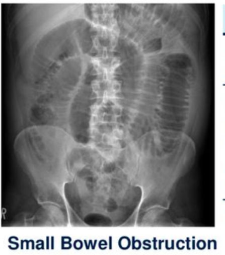 | Large bowel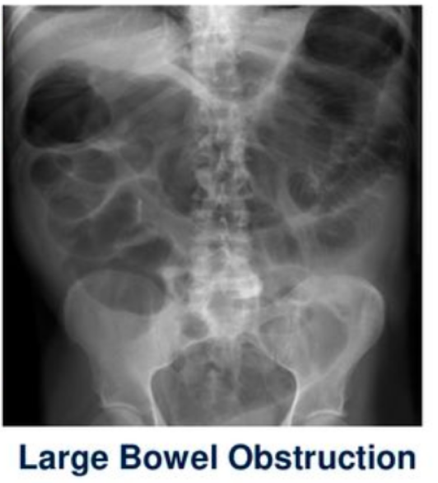 | |
| Location of dilated bowel loops | Central abdomen | Peripheral abdomen |
| Bowel wall pattern | Valvulae conniventes – folds/lines that cross full width of bowel | Haustral folds (lines do not cross entire lumen) |
| Air-fluid levels (Erect film) | Multiple short, “step-ladder” – like air-fluid levels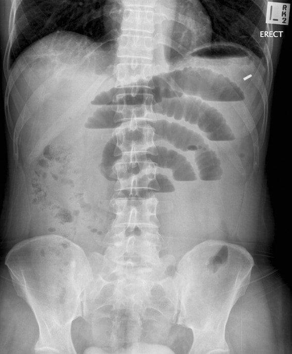 (image from https://litfl.com/axr-interpretation/) | Fewer, longer air-fluid levels |
| Gas and distension pattern (supine) | Proximal distension + no colonic gas | Proximal distension + distal decompression No small bowel air if competent ileocaecal valve |
| Thumbprinting | May be present due to bowel wall oedema/ischaemia image from https://www.radiologymasterclass.co.uk/tutorials/abdo/abdomen_x-ray_abnormalities/pathology_inflammatory_bowel | |
| Pneumoperitoneum (erect/decubitus) | Seen as free air under diaphragm if perforation occurs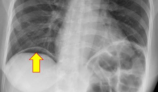 | |
| Side note NORMAL X-RAY DIFFERENCES OF SMALL AND LARGE BOWEL | ||
| Small bowel | Large bowel | |
| Location | Central | Peripheral |
| Content | Fluid and air | Faecal matter |
| Wall Patern | Valvulae conniventes (transverse mucosal folds that cross entire lumen) | Haustral folds (do not cross entire lumen); interspersed with plicae semilunares |
| Size | 3cm diameter | 6cm diameter (caecum 9cm) |
| Remember: 3, 6, 9 Rule |
| Small intestine 3cmLarge intestine 6cmCaecum 9cm |
| Remember Any increase in these numbers signify dilatation most likely due to an obstruction. |
Laboratory investigations
Additional investigations are done to investigate potential underlying causes or determine the presence of ischaemic changes3
- FBC: Leucocytosis
- ABG including lactate
- Metabolic acidosis and ↑lactate if ischaemic
- Metabolic alkalosis in dehydration and vomiting
- UEC
- hypokalaemia in patients who are severely dehydrated and vomiting
- HYPERkalaemia in ischaemia
- ↑urea:creatinine —> dehydration and AKI
- CRP elevated
Differential diagnosis
- For mechanical bowel obstruction:
- Paralytic ileus
- Acute or chronic mesenteric ischaemia
- IBD
- For small bowel obstruction:
- Appendicitis
- Pancreatitis
- For large bowel obstruction:
- Diverticulitis
- Toxic megacolon
- Ogilvie syndrome
Classification
Bowel obstructions can be classified in a variety of ways, such as:
- Anatomical location:
- Small or Large bowel
- Extent of obstruction
- Partial: Still can pass gas ± stool
- Complete: Unable to pass gas or stool (obstipation)
- Closed-loop obstruction: A form of complete bowel obstruction that occurs when a segment of the intestine is obstructed in TWO locations
- Presence of intestinal ischaemia:
- Mechanism of obstruction
- Mechanical obstruction: Volvulus, intussusception, colon cancer, hernia, adhesions
- Non-mechanical obstruction (pseudo-obstruction)
| Remember [Pseudo-obstruction refers to intestinal dysmotility syndromes that have signs, symptoms, and the radiologic appearance of obstruction in the absence of a mechanical cause.] |
- Ileus: A temporary paralysis of the myenteric plexus, leading to impaired transit of intestinal contents In the absence of mechanical obstruction. It typically presents acutely with severe pain, nausea and vomiting, distension and the inability to pass stool or tolerate food. Mostly resolves spontaneously
- Toxic Megacolon: A life-threatening dilation of the colon, often due to inflammatory or infectious causes. It can arise from Hirschsprung’s disease, idiopathic chronic constipation, or generalized GI dysmotility (intestinoparesis).
- Ogilvie’s Syndrome: Also known as acute colonic pseudo-obstruction, it presents as massive right-sided colon dilation without a mechanical cause, typically in hospitalised, medically unwell, or post-surgical patients.
- Hirschsprung’s disease: A paediatric condition where parts of the colon are aganglionic and lack the necessary innervation to contract, resulting in a non-mechanical obstruction. It is the most common cause of congenital megarectum and megacolon
Treatment
1. Initial Supportive Management (for ALL patients):5
- Resuscitation and Pre-op prep
- ABCDE
- IV fluids
- IV Analgesia
- Nil by mouth
- IV antibiotics: Indicated if suspected bowel compromise (ischaemia, necrosis or perforation)
- Remove/discontinue possible causative agents (in the case of medication induced ileus)
- Nasogastric (NG) Tube for Bowel Decompression:
- Begin gastric decompression to relieve pressure and reduce distension.
- The NG tube can also assist with feeding contrast for imaging or bypassing the obstruction temporarily if clinically indicated.
2. Definitive Management (if complications are present):5
- Urgent Surgical Intervention is Required If:
- There are signs of bowel ischaemia, perforation, or peritonitis.
- Surgical Options Include:
- Emergency laparotomy with procedures such as colectomy or Hartmann’s procedure, depending on the extent and location of pathology.
- Address the underlying cause directly (e.g., resection of tumour, lysis of adhesions, repair of hernia).
Complications and Prognosis
Complications
- Cardiovascular
- Hypotension
- Gastrointestinal
- Bowel ischaemia, perforation and peritonitis
- Systemic:
- Sepsis from perforation
Prognosis
Prognosis varies based on cause and timeliness of intervention. Most mechanical obstructions require surgery if complications arise. Delayed treatment increases risk of ischaemia, sepsis, and death. Functional obstructions (e.g., ileus) typically resolve with supportive care.