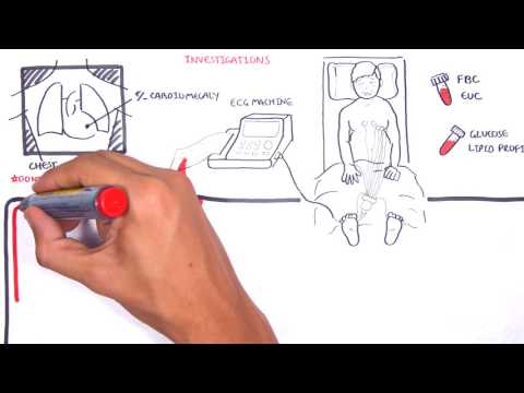
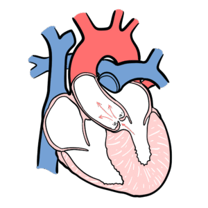
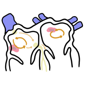
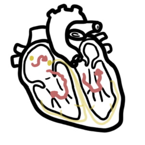
0:00 Hello, in this video we're going to talk about the anatomy of the heart, just 0:09 briefly. 0:10 So before we continue, just remember that the blue eye draw represents deoxygen 0:16 ated blood 0:17 and the red represents oxygenated blood, so blood high in oxygen. 0:22 So here we're looking at the anterior view of the heart, the superficial 0:26 anatomy. 0:27 The superior vena cava and the inferior vena cava bring deoxygenated blood to 0:31 the right 0:31 side of the heart, to the right atrium, then to the right ventricle. 0:35 The right ventricle will pump this deoxygenated blood up to pulmonary trunk and 0:40 then into 0:40 the left pulmonary artery and then into the right pulmonary artery. 0:46 The left and right pulmonary artery will bring this blood to the lungs and then 0:50 the blood 0:51 from the lungs will return to the heart to the left side of the heart via the 0:55 left pulmonary 0:57 vein and the right pulmonary vein. 0:59 You'll go to the left atrium here and then the left ventricle here and then 1:04 from the 1:05 left ventricle up to the aorta. 1:09 And the top part of the aorta here is known as your aortic arch. 1:13 The blood can then go up to the upper body or down to the lower body via the 1:17 descending 1:17 aorta from the back. 1:21 Now there is a ligament here that connects the aortic arch with the pulmonary 1:25 trunk. 1:25 This is a ligamentum arteriosum which is a remnant of the patent up this arter 1:30 iosus. 1:31 Here's the apex of the heart. 1:34 So that was the anterior view. 1:35 Let us look at the posterior view just to see where we are. 1:39 Here is again the superior vena cava and the inferior vena cava which will 1:43 bring deoxygenated 1:44 blood back to the right side of the heart, to the right atrium, then to the 1:49 right ventricle 1:50 here. 1:52 The right ventricle will then pump the blood up through the pulmonary trunk 1:56 here and then 1:56 from the pulmonary trunk it will bifurcate to the left and right pulmonary 2:02 artery. 2:03 The blood goes to the lungs and then return back to the side to the left side 2:07 of the heart 2:07 via the left and right pulmonary veins. 2:12 The left and right pulmonary veins enters the left atrium, left the left atrium 2:17 then 2:17 goes to the left ventricle which then goes and pumps this blood up the aorta. 2:22 Here is your aortic arch and then it can pump it up to the upper body or down 2:26 through 2:26 the descending aorta. 2:30 So now I want to cut a cross section of the anterior view of the heart and see 2:33 what's 2:34 happening inside. 2:36 And straight away you can see that there are four chambers within the heart. 2:42 And these are your atrium and your ventricles. 2:45 So just again recapping, here's your superior vena cava and inferior vena cava 2:50 which will 2:50 bring blood to the right side of the heart, to the right atrium. 2:54 From the right atrium blood flows to the right ventricle through a valve known 2:59 as a tricuspid 3:00 valve. 3:01 And then from the right ventricle blood will flow up to the pulmonary trunk 3:06 through another 3:06 valve called the pulmonary valve. 3:09 From the pulmonary trunk blood will go to the lungs. 3:13 From the lungs blood will return to the left side of the heart via the 3:17 pulmonary veins 3:18 into the left atrium. 3:20 From the left atrium blood flows to the left ventricle via the mitral valve. 3:27 From the left ventricle the left ventricle pumped this blood up the aorta via 3:32 the aortic 3:33 valve. 3:34 And then blood will flow up to the upper body or down to the lower body via the 3:38 descending 3:39 aorta. 3:41 Here is the apex of the heart again. 3:47 Now that we know the flow of blood through the heart, let us look at the flow 3:52 of blood 3:52 from the heart to the body tissues and also from the heart to the lungs in a 3:58 bit more 3:58 detail. 3:59 This is your systemic and pulmonary circulation. 4:05 So here I'm drawing the heart and here is your lower body and upper body. 4:10 So once your tissues around your body use oxygen, deoxygenated blood will 4:14 return to the right 4:16 side of the heart. 4:17 So here from the upper body blood will return to the heart and from the lower 4:22 body blood 4:22 will return to the right side of the heart to the right atrium and then to the 4:26 right 4:27 ventricle. 4:28 The deoxygenated blood will be pumped through the pulmonary trunk and towards 4:32 the lung via 4:33 the right and left pulmonary arteries. 4:37 Now here I'm drawing the right lung and the left lung. 4:40 Now this deoxygenated blood will undergo gas exchange within the lungs, 4:45 represented in 4:46 orange. 4:47 So what happens here is that carbon dioxide will be released and the lungs will 4:53 reoxygenate 4:54 the blood with oxygen and so now there is reoxygenated blood or oxygenated 5:00 blood returning 5:01 to the heart to the left side of the heart. 5:04 Then in the lungs, carbon dioxide is removed and the lungs will reoxygenate the 5:09 blood and 5:10 so now in red, the oxygenated blood will return to the left side of the heart 5:14 to the 5:14 left atrium and then to the left ventricle and then from the left ventricle, 5:18 the blood 5:19 is pumped up the aorta to the upper body or to the lower body. 5:24 And again, this oxygen will be delivered to the tissues here and again, gas 5:30 exchange 5:30 occurs represented in orange. 5:32 What happens is that oxygen is offloaded to the body tissues and then as a by 5:38 product, 5:39 carbon dioxide is released back into the blood and so you get deoxygenated 5:43 blood once again 5:44 returning to the right side of the heart. 5:48 Here again, you have oxygen being offloaded to the tissues and as a byproduct, 5:52 carbon 5:53 dioxide is released and then you have deoxygenated blood returning to the right 5:57 side of the heart 5:58 and the cycle continues. 6:00 This diagram represents both pulmonary circulation and systemic circulation. 6:08 Now that we know the lungs and the heart are in very close proximity, I wanted 6:12 to look 6:13 at something clinical looking at a chest x-ray and looking at each part of the 6:18 x-ray represents. 6:20 So coming off the left here is a normal chest x-ray and the area is represented 6:27 here. 6:27 So this is your right lung and left lung and here is your superior vena caper, 6:31 not your 6:33 IVC or SVC. 6:35 Here's your right atrium and here is your right ventricle. 6:38 This is your pulmonary trunk here. 6:40 Here's your left atrium and left ventricle and this is your aorta. 6:44 There are important angles to remember when looking at a chest x-ray. 6:48 These are your right cardiophonic angles and your left cardiophonic angles and 6:53 then your 6:53 right costophonic angles and your left costophonic angles. 6:58 These areas are important because blunting of these angles will represent 7:02 something 7:02 called pleural effusion. 7:05 Another important landmark to remember are your right and left clavicles here. 7:10 And in the middle you can see tracheal shadows. 7:13 The tracheal shadowing is really important because deviation of the tracheal 7:18 shadow to 7:18 the left or right can signify something called attention pneumothorax. 7:24 So again this diagram that I drew represents a normal chest x-ray and it's 7:29 important to 7:30 remember some of these areas because you can compare it to an ab normal chest x 7:35 -ray such 7:35 as this one on the right which is a chest x-ray of someone who has heart 7:40 failure. 7:41 And straight away you can see that the heart is big. 7:44 It occupies more than 50% of the cardiothoracic ratio and straight away you can 7:49 see partly 7:50 blunting of some of the angles, the cardiophrenic and costophrenic angles. 7:56 So I hope this video was helpful, this was basic anatomy of the heart. 7:59 You can continue watching clinical anatomy of the heart focusing on the valves, 8:05 the pericardium 8:06 and also the coronary arteries and also the coronary vessels. 8:11 Thank you for watching.