

Mitral Regurgitation (Incompetence)
Overview
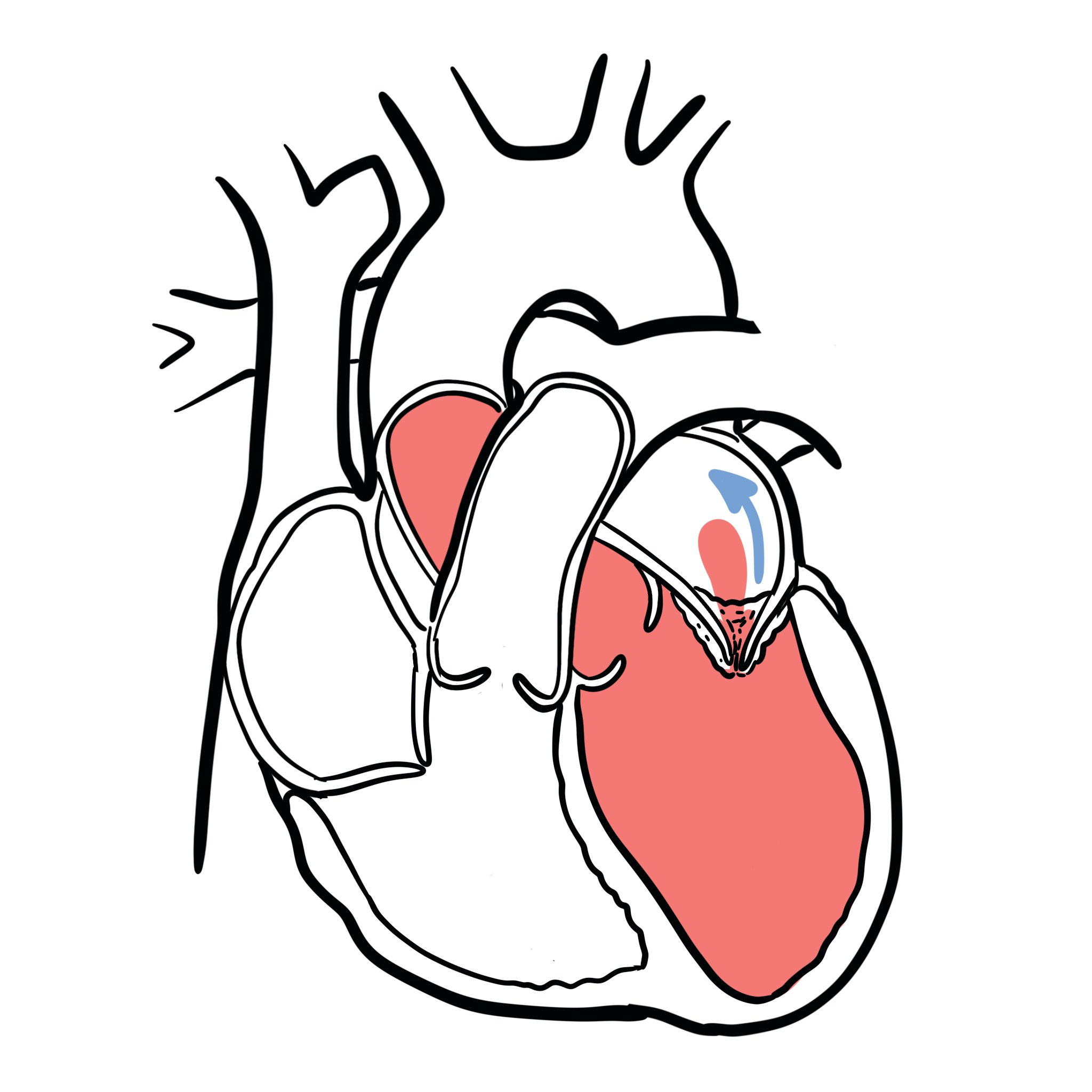
Mitral regurgitation (MR), which is also known as mitral insufficiency, is a common heart valve disorder. When MR is present, blood leaks backwards through the mitral valve when the heart contracts. This reduces the amount of blood that is pumped out to the body. Patients with acute mitral regurgitation (MR) are often gravely ill with significant haemodynamic abnormalities that require urgent medical and usually surgical treatment.
Definition
Primary MR: intrinsic disease of the valve leaflets, chordae, papillary muscles or annulus.
Secondary MR: valve leakage caused by left-ventricular or left-atrial remodelling rather than primary leaflet disease.
Mitral valve prolapse: displacement of one or both mitral leaflets into the left atrium during systole.
Flail leaflet: loss of leaflet support, usually due to chordal rupture, causing the leaflet edge to move freely into the left atrium.
Transcatheter edge-to-edge repair—TEER: catheter-based approximation of the mitral leaflets to reduce regurgitation.
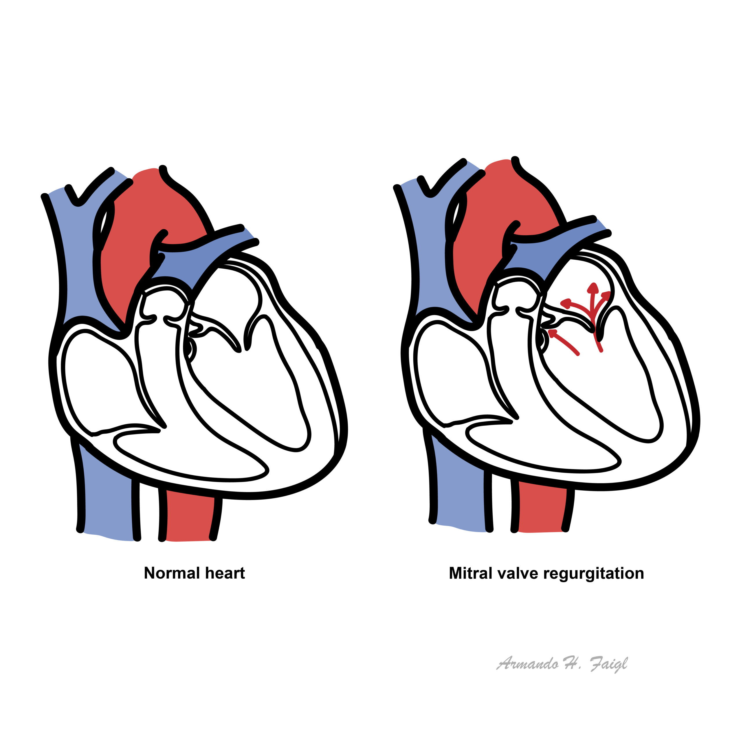
What to notice
Detailed image description
MR can be acute (pulmonary oedema), chronic compensated or chronic decompensated.
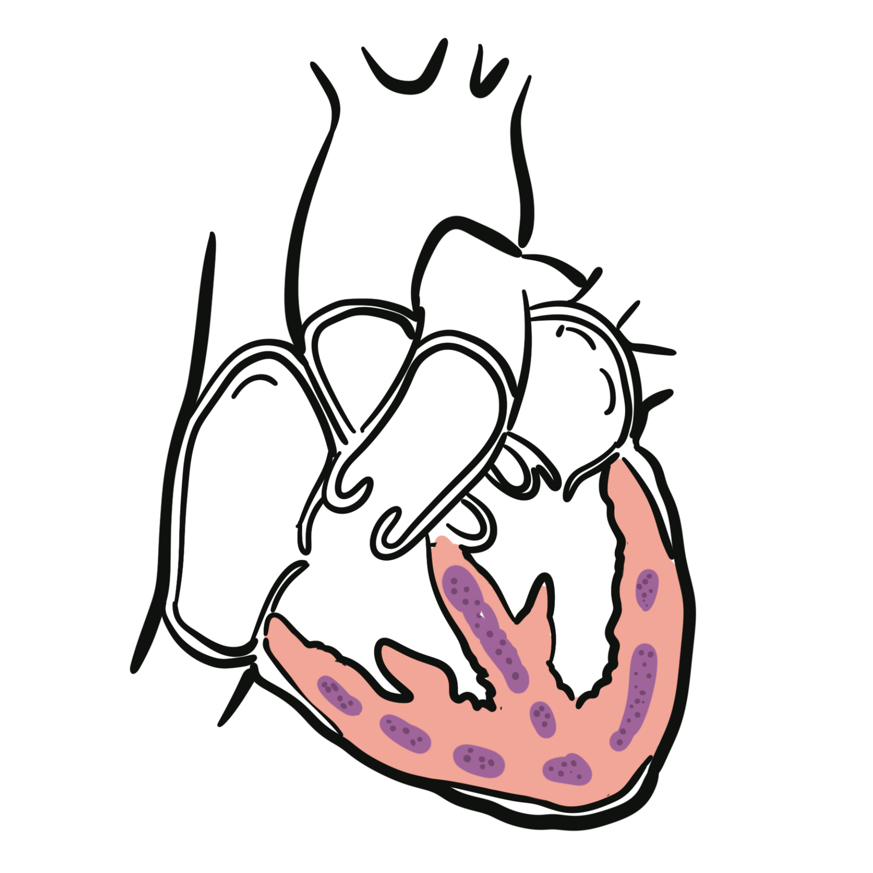
Anatomy
Heart Valve
The heart has 4 valves:
- Aortic valve
- Pulmonary valve
- Tricuspid valve
- Mitral valve (bicuspid valve)
The Mitral and tricuspid valves are atrioventricular valves meaning they allow blood to move from the atrium to the ventricles of the heart. This occurs with ventricular diastole.
The aortic and pulmonary valve are tricuspid valves which when open allow blood to move to the aorta and pulmonary system respectively. This occurs during ventricular systole when the heart contracts.
Surface anatomy of the heart valves
All of the heart valves except the mitral valve are usually tricuspid. However, there can be congenital bicuspid valves which can predispose one to valvular disease later on. The heart valves can be heard most prominent in the following regions of the chest
- Aortic valve – Right 2nd intercostal space parasternal
- Pulmonary valve – Left 2nd intercostal space parasternal
- Tricuspid valve – Left 4th intercostal space parasternal
- Mitral valve – Left 5 intercostal space mid-clavicular (below the nipple)
Aetiology & Risk Factors
Aetiology
There are three basic mechanisms of Mitral regurgitation:
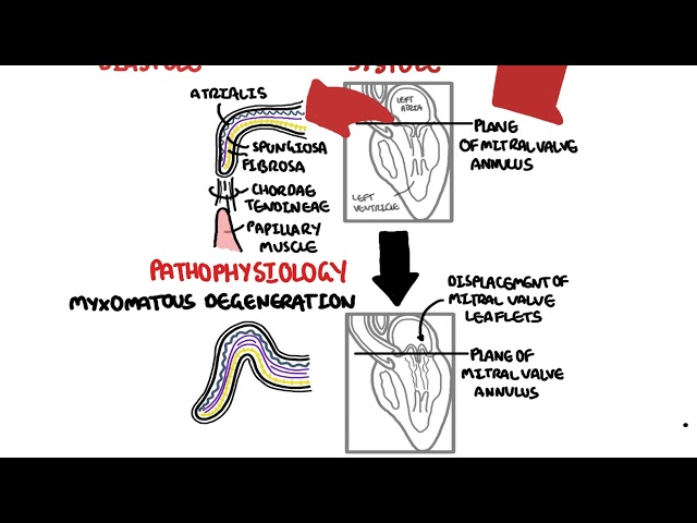
- Ruptured mitral chordae tendineae (flail leaflet) due to myxomatous disease (mitral valve prolapse), infective endocarditis, trauma, rheumatic heart disease (acute rheumatic fever or chronic rheumatic mitral valve disease), or spontaneous rupture.
- Papillary muscle rupture due to acute myocardial infarction or trauma or papillary muscle displacement due to myocardial infarction or ischemia.
- Induction of MR in the setting of dynamic left ventricular outflow obstruction. This complication has been observed in patients with left ventricular hypertrophy (including patients with hypertrophic cardiomyopathy), as well as in patients with myocardial infarction or Takotsubo cardiomyopathy
Another way of thinking about aetiology. Primary causes include abnormality of dysfunction of any component of the mitral valve apparatus. Secondary causes (functional MR) abnormality of dysfunction of the left ventricle.
| Risk Factors |
| Mitral valve prolapse |
| History of rheumatic heart disease |
| Infective endocarditis |
| History of cardiac trauma |
| History of ischemic heart disease/ MI |
| Congenital heart disease |
| Hypertrophic cardiomyopathy |
| Anorectic/dopaminergic drugs |
Classification
Primary – degenerative or organic – Mitral regurgitation
Caused by structural damage to the mitral-valve apparatus:
- Mitral valve prolapse
- Myxomatous degeneration
- Chordae tendineae rupture
- Flail leaflet
- Rheumatic heart disease
- Infective endocarditis
- Mitral annular calcification
- Congenital valve abnormalities
- Papillary-muscle rupture after myocardial infarction
Secondary – functional – Mitral regurgitation
The valve leaflets are usually structurally normal.
- Ventricular secondary MR:
- Ischaemic cardiomyopathy
- Dilated cardiomyopathy
- Left-ventricular dilatation and systolic dysfunction
- Papillary-muscle displacement and leaflet tethering
- Atrial secondary MR:
- Chronic atrial fibrillation
- Left-atrial and mitral-annular dilatation
- Often associated with heart failure with preserved ejection fraction.
Acute versus chronic Mitral regurgitation
- Acute MR: develops suddenly, such as after papillary-muscle rupture, chordal rupture or infective endocarditis.
- Chronic MR: develops gradually, allowing compensatory enlargement of the left atrium and ventricle.
Pathophysiology
Pathophysiology Acute MR
- Primary and secondary causes → weak mitral valve causing regurgitation → ↑preload and ↓afterload → ↑strokevolume
- ↑stroke volume → blood flows toe right atrium through incompetent mitral valve → pulmonary veins → pulmonary oedema
Pathophysiology Chronic MR
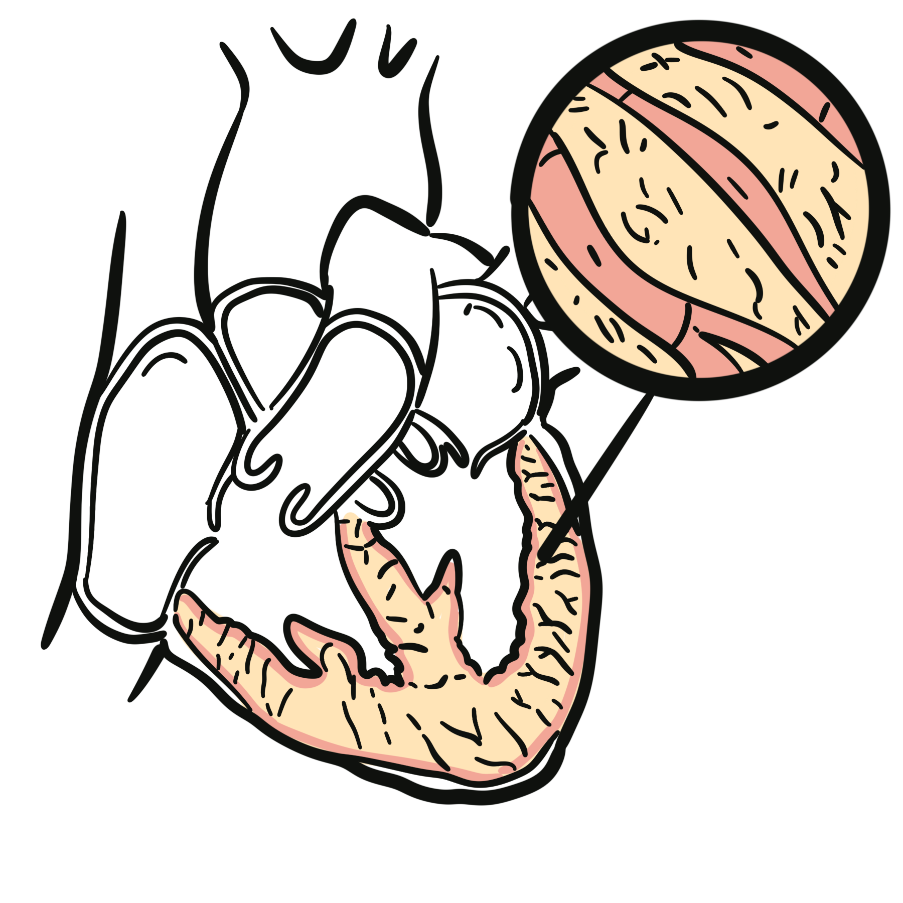
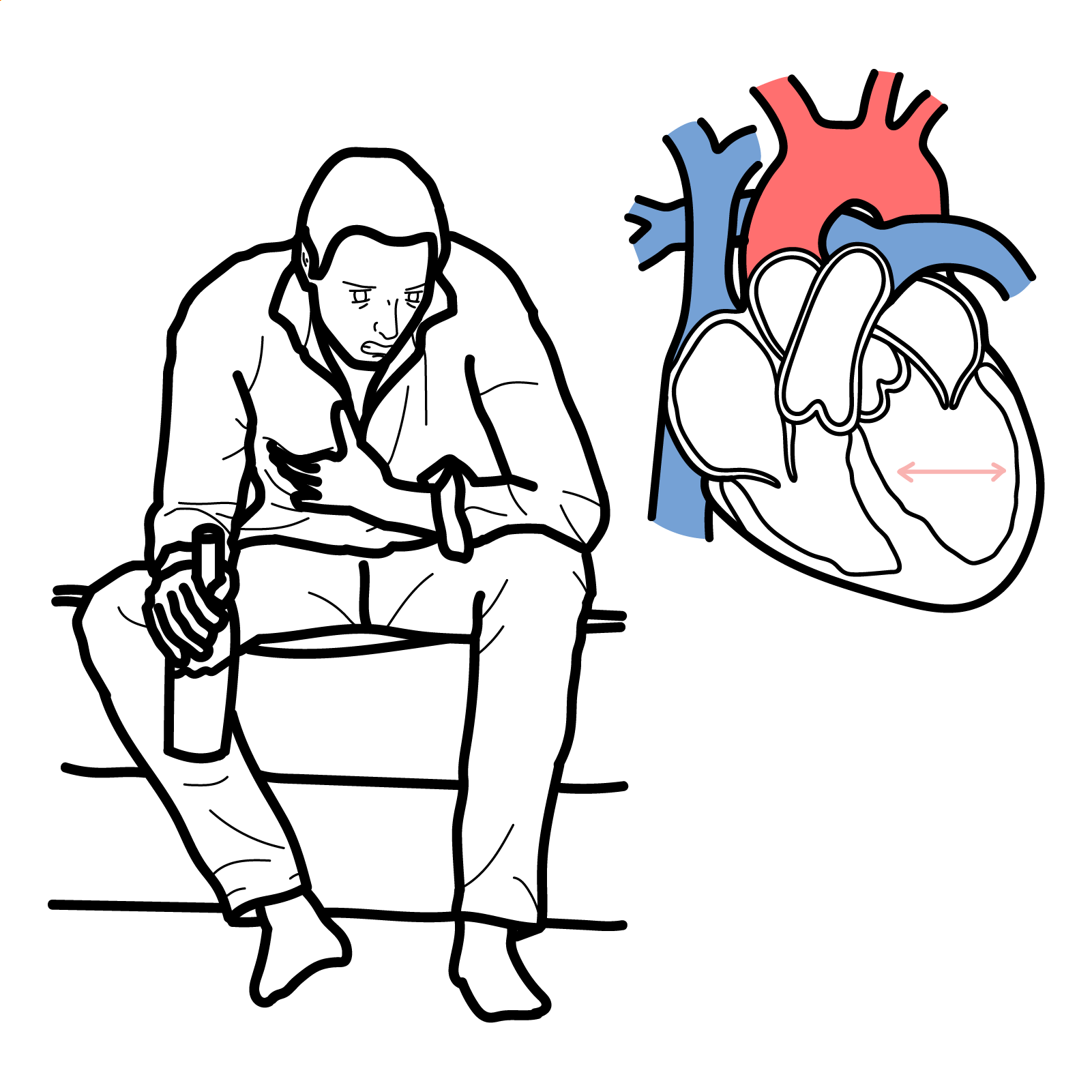
- Compensated Overtime the left ventricle will compensate for the↑preload → Enlargement of left ventricle → Dilated cardiomyopathy (eccentric left ventricular hypertophy)
- Decompensated Overtime the dilated cardiomyopathy gets bad until ejection fraction drops. Low ejection fraction occurs as a result of afterload acess and depressed myocardial contractility.
Clinical Manifestation
Acute MR presents as a cardiac emergency, however chronic MR typically presents with no symptoms until late stage.
Acute Mitral Regurgitation presents with Left sided heart failure.
- Pulmonary oedema
- Hypotension
- Cardiogenic shock
- Pallor
- Diaphoresis
- Tachycardia
- Signs of right sided heart failure
- Raised JVP
- Peripheral oedema
- Sign of AF
- palpitation
Most people with mitral regurgitation (MR) have no symptoms. People with mild to moderate MR may never develop symptoms or serious complications. A trivial amount of mitral regurgitation (MR) is present in up to 70 percent of adults. Significant (moderate to severe) MR is much less common.
A pansystolic murmur at the apex radiating to the axilla suggests MR. Acute severe MR may have a surprisingly soft murmur despite pulmonary oedema or shock.
Cardiac Examination
- Holosystolic (Pansystolic murmur) – occurs during systole
- Murmur radiates to axilla
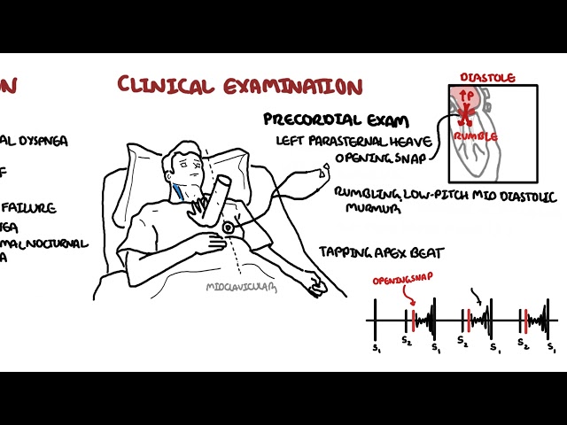
Cardiac Examination includes a murmur heard over the left 5th intercostal space mid-clavicular. The murmurs is a flat continuous murmur which radiates to the axilla. There also tends to be a diminished S1.
Clinical Signs of severity
- Enlarged left ventricle
- Pulmonary hypertension
- Left ventricular failure
- Third hard sound, S3
- Soft first heart sound
- S2 splitting (A2 heard before P2)
- Small volume pulse
Breathing out as if you were straining (vasalva manoevre) increases intrathoracic pressure -> decreases venous return and accentuates murmur from mitral valve prolapse and hypertrophic obstructive cardiomyopathy.
Diagnosis
Diagnosis
- Transthoracic echocardiography—first-line
- Transoesophageal echocardiography
Investigations
- Chest X-ray
- Exercise tolerance test
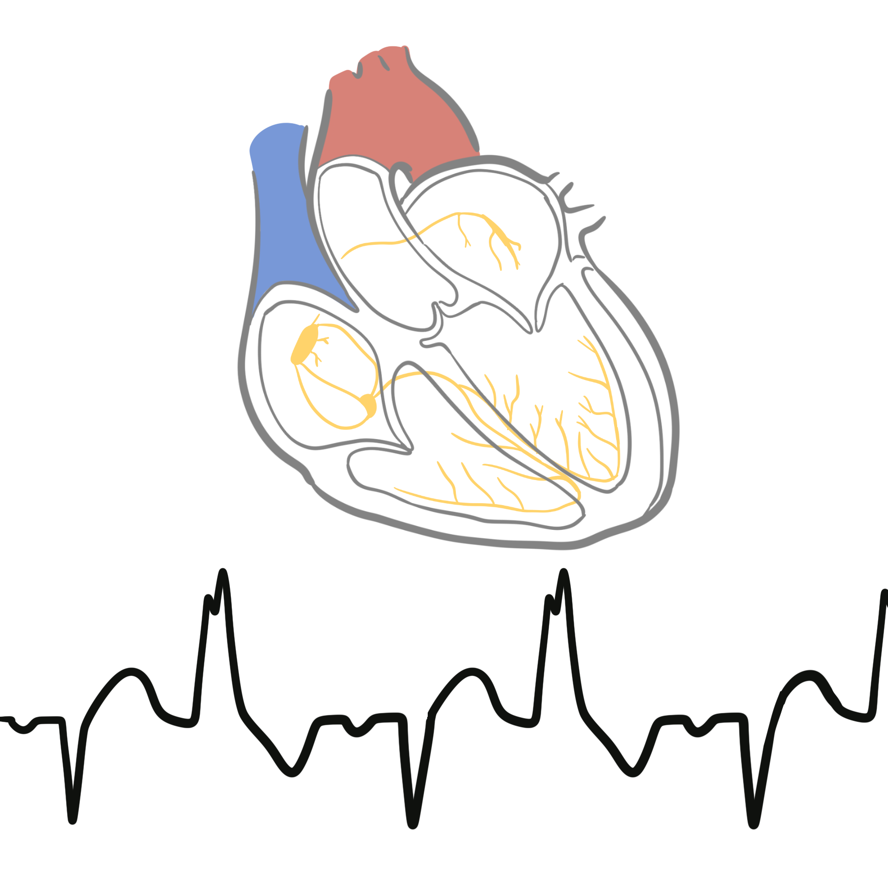
- ECG – detect rhythm disturbances or evidence of coronary heart disease or other conditions that can cause MR. It can also show evidence of other associated cardiac abnormalities.
- Exercise testing or stress echocardiography: useful when symptoms and resting MR severity are discordant.
- Cardiac MRI: accurately assesses ventricular volumes, regurgitant volume and myocardial fibrosis when echocardiography is inconclusive.
- Coronary angiography or CT coronary angiography: before valve surgery in patients with relevant coronary risk or suspected ischaemic disease.
- Right-heart catheterisation: considered when pulmonary pressures are uncertain or discordant with non-invasive assessment.
Differential Diagnosis
- Acute Coronary Syndrome
- Mitral stenosis
- Aortic stenosis
- Infective endocarditis
- Atrial Myxoma
Treatment
Acute severe Mitral Regurgitation
- Medical emergency requiring urgent cardiology and cardiothoracic assessment.
- Oxygen and ventilatory support as required.
- IV diuretics for pulmonary oedema.
- Vasodilator therapy may reduce afterload if blood pressure permits.
- Vasopressors or mechanical circulatory support may be required in shock.
- Treat the cause, such as myocardial infarction or infective endocarditis.
- Papillary-muscle rupture or other mechanical failure usually requires urgent surgical repair or valve replacement.
Always distinguish primary valve disease from secondary functional MR, because treatment differs.
Definitive management
Acute Mitral Regurgitation
Inoperable patients
- Valvuloplasty
- Annuloplasty
Operable patients
- Valve Replacement (~Ejection Fraction <60)
- mechanical
- bioprosthetic
Chronic Mitral Regurgitation
- ACE inhibitor or beta-blocker (Asymptomatic)
- Vasodilators
- Serial echocardiograms for monitoring
- Surgery as above
If in AF, anticoagulate with warfarin (however now NOACs are being more frequently used).
Complications & Prognosis
Complications
- Left-ventricular dilatation and systolic dysfunction
- Acute pulmonary oedema
- Atrial fibrillation
- Left-atrial thrombus and embolic stroke
- Pulmonary hypertension
- Right-sided heart failure
- Infective endocarditis
- Sudden cardiac death, particularly in advanced disease
- Progressive functional limitation and reduced quality of life.
- Treatment related complications
- Infection or prosthetic-valve endocarditis
- Residual or recurrent MR
- Mitral stenosis following repair
- Valve thrombosis or structural deterioration
- Need for permanent anticoagulation with a mechanical valve
Prognosis
- Mild MR may remain stable for many years.
- Severe untreated MR can lead to irreversible ventricular dysfunction, atrial fibrillation, pulmonary hypertension, heart failure and increased mortality.
- Successful durable surgical repair provides excellent long-term outcomes in appropriately selected patients.
- Prognosis in secondary MR is strongly influenced by the severity of the underlying cardiomyopathy, ventricular function, renal function, pulmonary hypertension and response to heart-failure therapy.
References
- Nishimura RA, O’Gara PT, Bavaria JE, et al. 2020 focused update of the 2017 ACC expert consensus decision pathway on the management of mitral regurgitation. J Am Coll Cardiol. 2020;75(17):2236–2270. doi:10.1016/j.jacc.2020.02.005.
- Praz F, Borger MA, Lanz J, et al. 2025 ESC/EACTS Guidelines for the management of valvular heart disease. Eur Heart J. 2025;46(44):4635–4736. doi:10.1093/eurheartj/ehaf194.
- Otto CM, Nishimura RA, Bonow RO, et al. 2020 ACC/AHA guideline for the management of patients with valvular heart disease. Circulation. 2021;143(5)–e227. doi:10.1161/CIR.0000000000000923.
- Zoghbi WA, Asch FM, Bruce C, et al. Guidelines for the evaluation of valvular regurgitation after percutaneous valve repair or replacement. J Am Soc Echocardiogr. 2019;32(4):431–475. doi:10.1016/j.echo.2019.01.003.
- Stone GW, Abraham WT, Lindenfeld J, et al. Five-year follow-up after transcatheter repair of secondary mitral regurgitation. N Engl J Med. 2023;388(22):2037–2048. doi:10.1056/NEJMoa2300213.








Members only discussions coming soon…