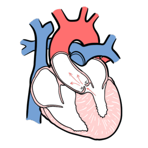
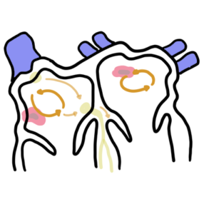
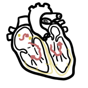
0:00 Mitral stenosis is a condition characterized by obstruction of blood flow 0:10 across the mitral 0:11 valve from the left atrium to the left ventricle. 0:14 The mechanical obstruction leads to increases in pressure within the left at 0:20 rium, pulmonary 0:21 vasculature and right side of the heart. 0:30 The mitral valve is an atrium ventricular valve, separating the atrium and the 0:35 ventricle. 0:36 The tricuspid valve is also an atrium ventricular valve. 0:40 The mitral valve is made up of two leaflets. 0:44 The normal area of the mitral valve is 4 to 6 centimeter squared. 0:52 During diastole, which is when the ventricles fill, the atrium ventricular 0:59 valve are open, 0:59 allowing the blood to flow from the atrium to the ventricle. 1:04 During ventricular systole, the mitral valve closes, allowing blood to be 1:11 ejected from 1:11 the aorta into circulation. 1:17 The normal cardiac and pulmonary circulation pressures are important to know. 1:23 As you can see within the aorta, which is the blood pressure pressure, is about 1:27 120 1:28 on 80 millimeters murky. 1:30 The left atrial pressure is 6. 1:37 The narrowing of the mitral valve, as seen in mitral stenosis, to less than 2.5 1:43 centimeter 1:44 squared, will impede the free flow of blood between the left atrium and the 1:50 left ventricle. 1:52 This causes an increase in left atrial pressure. 1:56 The increase in atrial pressure is required to maintain outflow and adequate 2:02 cardiac output 2:03 of the heart. 2:08 When the mitral valve becomes significantly narrow, less than 1 centimeter 2:12 squared, this 2:14 is critical stenosis, critical obstruction. 2:17 At this stage, the atrial pressure is at least 25 millimeters murky, in order 2:23 to maintain 2:24 that same cardiac output. 2:26 However, in mitral stenosis, as left atrial pressure increases, and the left at 2:32 rial continues 2:33 to dilate, blood will pull backwards, causing pulmonary hypertension. 2:41 You can imagine blood pulling from the left atrial to the pulmonary circulation 2:46 , causing 2:47 pulmonary hypertension, as well as pulmonary edema. 2:52 These changes mimic left-sided heart failure, due to the pulmonary edema and 2:57 pulmonary hypertension. 2:59 But in reality, the left side of the heart, at least the left ventricle, is 3:03 functioning 3:04 normally in mitral stenosis, and cardiac output can be maintained. 3:12 As left atrial pressure rises, pulmonary hypertension occurs, which can lead to 3:16 pulmonary regurgitation, 3:19 as well as tricuspid regurgitation, and then secondary, right-sided heart 3:25 failure. 3:27 Right-sided heart failure will cause pressure to increase in the venous 3:31 circulation. 3:33 So all that blood returning to the right side of the heart can pull backwards, 3:37 leading to 3:37 a tender, enlarged liver, and then subsequently, portal hypertension and asc 3:44 ites, and peripheral 3:46 edema. 3:50 Left atrial pressure and the dilatation of the left atrial specifically can 3:55 contribute 3:56 to atrial fibrillation, as well as stasis of blood in the area. 4:02 Stasis of blood increases the risk of thrombus formation and then systemic emb 4:07 oli, which 4:08 can lead to a stroke. 4:14 The etiology of mitral stenosis is many, however, rheumatic fever is the most 4:21 common cause, 4:22 which will then can lead to rheumatic heart disease. 4:27 Rheumatic fever is caused by streptococcus pyogenes strep throat. 4:32 Antibodies are then produced, however, unfortunately, there is molecular mimic 4:36 ry between the heart 4:37 valves and the M protein of strep pyogenes, and this causes a cross-reaction 4:42 and subsequent 4:43 rheumatic heart disease. 4:48 Carcinoid syndrome is another cause, and this is due to carcinoid tumours, 4:52 which is normally 4:53 associated with tricuspid valve disease. 4:58 Congenital mitral stenosis is another cause, as well as rheumatoid arthritis 5:02 and systemic 5:03 lupus erythmatosis. 5:06 Other rare causes include Fabry's disease, a rare inherited disease caused by 5:11 deficiency 5:12 of an enzyme called alpha-galactocidase-A. 5:18 Whipple's disease, a rare systemic infectious disease caused by the bacterium 5:22 tropharema 5:23 whipply, and this can cause vegetations in the mitral valve leading to stenosis 5:34 . 5:35 The clinical manifestations of mitral stenosis, mitral stenosis usually 5:39 presents with exertional 5:41 dyspnea, and/or a decrease in exercise tolerance. 5:45 There can be atrial fibrillation, which would present as palpitation or a throm 5:49 botic event, 5:50 such as a stroke. 5:53 The person may also have symptoms of left-sided heart failure, orthopnea, parox 5:57 ysmal dyspnea, 5:58 and fatigue. 6:00 Right-sided heart failure symptoms in signs is more specific to mitral stenosis 6:05 and includes 6:06 peripheral edema. 6:11 Clinical examination. 6:13 In the precordial exam, there is a tapping apex beat in the fifth intercostal 6:18 space 6:18 in the mid-clavicular line. 6:20 There can be a left-parastonal heave, which indicates right ventricular enlarg 6:25 ement. 6:26 On oscotation, you can hear an opening snap, and this is caused by opening of 6:30 the stenosis 6:31 mitral valve, and indicates that the leaflets are pliable. 6:37 Following the opening, there's rumbling, low-pitch, mid-diastolic murmur. 6:41 This is heard best in the left lateral position on expiration. 6:45 The murmur is louder by increasing flow across the valve, so performing sit-ups 6:51 or jumps. 6:53 Also, there's a loud, first hot sound, which is the closing of the atrovent 6:59 ricular valve. 7:08 Remember signs of pulmonary hypertension, a loud P2 over the pulmonary valve, 7:12 and right-sided 7:14 heart failure. 7:16 So there is a distended neck veins, increased jugular venous pressure, there 7:20 can be asides 7:21 as well as peripheral edema. 7:26 Remember also signs of left-side heart failure, there may be coarse crackles. 7:32 Malaflush is caused by reduced cardiac output state, with associate vasod 7:36 ilation in the face. 7:43 Clinical signs, or clinical markers of severe much stenosis, include an earlier 7:47 opening 7:48 snap, increasing length of the murmur itself, signs of pulmonary hypertension, 7:54 pulmonary 7:54 edema, something called Graham's steel murmur, which is a sign of pulmonary reg 8:01 urgitation, 8:02 low pulse pressure. 8:05 When someone presents with what looks like a malaflush, think about other 8:08 differential 8:09 diagnoses as well, including hypothyroidism, cold weather can cause vasod 8:13 ilation in the 8:14 face, carcinoid syndrome, systemic lupus erythmatosis, causing the lupus 8:20 butterfly rash and polycythemia. 8:25 The differential diagnosis of a mitral stenosis murmur, so what we're talking 8:29 about is that 8:30 mid-diastolic rumbling murmur, are three things. 8:35 The first is severe mitral annular calcification, which causes functional mit 8:41 ral stenosis due 8:42 to a reduction in the annular size and fibrocalcific changes of the mitral 8:48 leaflets, left atrial 8:50 mixoma, which is a benign cardiac tumour, that occurs most frequently in the 8:55 left atria 8:56 and can present with symptoms similar to mitral stenosis. 9:01 And the last is core triatum, which is division of the left or right atria by a 9:08 membrane that 9:09 may cause obstruction to flow. 9:16 And so investigations for suspected mitral stenosis, an ECG will show bifid p- 9:22 waves, which indicates 9:23 left atrial enlargement or dilatation, an ECG may also show atrial fibrillation 9:29 , an 9:29 irregular irregular heart rhythm. 9:33 A chest x-ray may show a double right heart border, which indicates an enlarged 9:39 left atrial 9:40 size, there may also be pulmonary congestion and prominent pulmonary arteries, 9:46 indicating 9:46 pulmonary hypertension. 9:49 Echocardiogram is for diagnosis, as well as to assess severity of mitral sten 9:55 osis. 9:56 Coronary angiogram can also be used to evaluate for coronary artery disease. 10:06 One of mitral stenosis includes pharmacotherapy and there are four main 10:10 medications. 10:11 The first is anticoagulants, including warfarin. 10:15 Warfarin is the anticoagulant of choice for patients who have mitral stenosis, 10:19 and this 10:20 is specifically to manage atrial fibrillation, as well as reduce the risk of 10:24 systemic embolization. 10:28 Diuretics, such as frouzamide to manage fluid overload, antirrhythmic agents in 10:32 the setting 10:33 of atrial fibrillation. 10:35 And finally, antibiotics, specifically prophylactic antibiotics, to reduce the 10:40 risk of infective 10:41 endocarditis, or joint infections in those who have a background of rheumatic 10:48 fever. 10:48 Interventions for mitral stenosis include percutaneous valvuloplasty, where 10:53 they essentially put 10:54 a balloon in between the mitral valve to dilate it. 11:00 So in summary, mitral stenosis is where you have obstruction of blood flow from 11:17 the left 11:18 atrium to the left ventricle. 11:20 It causes a mid-diastolic murmur, and the main cause is rheumatic heart disease 11:26 . 11:26 Thank you. 11:30 for watching. 11:31 [end of transcript]