Bone Lump (Bone Tumour)


Benign bone tumours account for 99% of cases. Malignant bone tumours account for 1% of cases.
History
Persistent deep pain not relieved with NSAIDs suggest malignant bone lump.
Examination
| X-ray Findings | |
| Benign | Malignancy |
| Regular border | Irregular border |
| Smaller size | Larger size |
| – | Codman’s Triangle |
| No sclerosis | Sclerosis |
| Homogenous | Heterogenousity |
| Non-invasive | Invasion |
Staging Investigations
Benign Bone Growth – Child or young adult without symptoms or with signs and symptoms of pain, swelling, decreased range of motion, deformity and pathological fracture.
Malignant Bone Growth
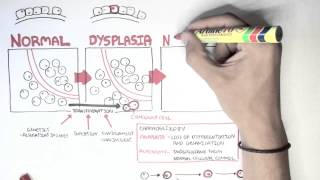
Malignant tumour: haemopoetic (common), metastatic bone tumour (very uncommon) and primary bone tumour (very uncommon).
| Benign tumour | Clinical Features | Origin | Pathology | Radiology |
| Osteoid osteoma | ||||
| Osteochondroma | Symptomatic: Pain, decreased range of motion, deformity | Metaphysis of long bones and the ileal crest | Benign cartilage-capped tumour attached to the underlying skeleton by a bony stalk.
| Well-defined bony protuberance emerging from metaphysis. Bony spur points away from the joint |
| Chondroblastoma | Low grade joint pain (constant, unrelated to activity) Swelling/joint effusion | Epiphysis of the proximal long bones | Composed of chondroblasts and chondroid matrix. There may be calcium deposition surrounding the chondroblasts causing a “chicken-wire calcification”. | Well-demarcated radiolucent area. Sclerotic border |
| Chondromyxoid fibroma | Pain, swelling, pathological fracture | Metaphysis of the bones of the lower limb | Solid tumour of mixed cartilage, fibrous and myxomatous (connective) tissues. | Eccentric, intramedullary, lobulated or bubbly lesion Sclerotic border Typically lucent |
| Giant cell tumour | Pain, swelling, decreased range of motion, deformity, pathological fracture | Epiphysis and diaphysis of long bones | Macroscopic – fleshy, reddish tumour often containing cystic and haemorrhagic areas. Tumour may breach the cortex and extend into the soft tissues. There is little to no periosteal reaction. Microscopic – abundance of giant cells scattered on a background of stromal cells. | Expansile, eccentrically-placed lytic area May extend into the joint No matrix calcification or periosteal new bone formation |
| Bone Cysts | Pain, swelling, decreased range of motion, deformity, pathological fracture | Metaphysis and diaphysis of long bones | Fluid filled lesions with a fibrous lining | Well-marginated cystic lesions (radiolucent) i.e. “large bubble inside the bone”. No sclerosis Fallen leaf sign – where area of pathological fracture falls to the bottom of the cyst |
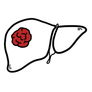
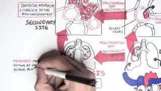
Giant cell tumours may metastasise to lungs but metastatic disease does not lead to patient death.
| Malignant tumour | Clinical Features | Origin | Pathology | Radiology |
| Osteosarcoma | Localised pain – deep, dull, boring and relentless. Resistant to analgesia Progressively enlarging mass tender to palpation Inflamed overlying tissue Pathological fracture | Metaphyseal region of long bones. 50% occur in knee (distal femur or proximal tibia). | Highly malignant tumour which arises within the bone and spreads rapidly outwards to the periosteum and soft tissues. Osteosarcomas are bulky tumours which are gritty, grey-white and often contain areas of haemorrhage and cystic degeneration. | Large destructive mixed lytic and blastic mass with infiltrative margins Codman triangle Sunburst appearance Poorly defined tumour margins |
| Chondrosarcoma | Age: Adults >40yo Localised pain – deep, dull, boring and relentless. Resistant to analgesia Progressively enlarging mass tender to palpation Inflamed overlying tissue Pathological fracture | Metaphysis of the proximal femur and humerus, pelvis, shoulder girdle and ribs. Can occur in any bone which develops in cartilage. | Malignant tumour composed of hyaline or myxoid cartilage. Large bulky tumours with grey/white or translucent nodules of cartilage. Spotty calcification Cystic spaces Thickened cortex | Mixed radiolucent and sclerotic appearance with mineralised chondroid matrix Thickened cortex No periosteal reaction |
| Ewing Sarcoma | Painful enlarging masses Tender to palpation Localised swelling and erythema Systemic findings mimicking infection | Diaphysis or metaphysis of bones of lower limb | Arises in medullary cavity and invades the cortex, periosteum and soft tissue. Tumour is soft, tan-white and may contain areas of haemorrhage and necrosis. Characteristic periosteal reaction produces layers of reactive bone | Destructive lytic tumour with permeative margins which extend into surrounding soft tissue. “Moth eaten” appearance “Onion peel” due to characteristic periosteal reaction |
Pain associated with malignant tumours: nocturnal pain, sudden severe pain – possible pathologic fracture, persistent pain, especially in children and pain which doesn’t respond to analgesia.
Codman triangle – triangular shadow between cortex and raised ends of periosteum due to tumour breaking through the cortex, lifting the periosteum and causing reactive periosteal bone formation.
Treatment of Malignant Tumours – depending on severity and type on bone tumours.


Discussion