Splenomegaly generally denotes a palpably enlarged spleen. Splenomegaly can be found in 3% of the normal population. Causes of an enlarged spleen are multiple (hepatic, haemotological, infection or inflammation).
Definition Hypersplenomegaly: abnormal enlargement of the spleen Hypersplenism: defined as one or more blood cytopenias in the setting of splenomegaly. Pancytopaenia: reduction in the number of RBCs, WBCs, and platelets in the peripheral blood below the lower limits of the age-adjusted normal range for healthy people. It is therefore the combination of anaemia, leukopenia, and thrombocytopenia.
Anatomy of the spleen
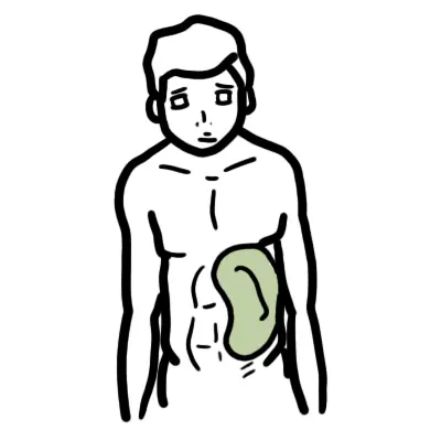
Location: Left hypochondrium
Rule of odds (1,3,5,7,9-11):
1 inch thick
3 inches broad
5 inches long
7 ounces weight
Underlies 9-11 ribs
Position: obliquely along long axis of 10th rib; directed downward, forward and laterally
Arterial supply: Splenic artery from celiac trunk
Venous drainage: Splenic vein → Portal vein
Lymphatic drainage: Celiac (Para-aortic) nodes
Nerve supply: Sympathetic from celiac plexus
Histology of the spleen
Red pulp: sinuses lined by endothelial macrophages and cords (spaces)
White pulp: structure similar to lymphoid follicles
Physiology of the spleen
Red pulp
White pulp
Removal of antibody-coated bacteria and antibody coated blood cells from circulation
Rashes and or joint swelling - Systemic autoimmune disease
Petechiae and ecchymoses - thrombocytopaenia , malignant haemotolgic disease
Side note Proper examination of the spleen requires relaxation of both the abdominal musculature (arms at the side of abdomen) of the patient and the examiner hands (beware of tender spleen)
Difference Between Enlarged Spleen and Kidney Examination
Enlarged Spleen
Kidney
Sharpe edge
Round edge
Crosses midline
Does not cross midline
Moves with respiration
Does not move with respiration
Cannet get above it
Can get above it
Approach in Adults
History
Recent trauma
Race
Fever
Recent infection
Recent travel
Constitutional signs - systemic illness including malignancy and autoimmune disease
Pruritis - Myeloproliferative disorder
Alcohol or hepatitis - portal hypertension secondary to liver disease
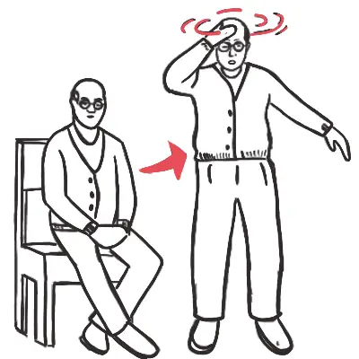
Symptoms of splenomegaly - Satiety, abdominal discomfort, dragging sensation, abdominal pain, shoulder tip pain
Remember symptoms of splenomegaly include early satiety, abdominal discomfort and pain, irritation of diaphragm may cause shoulder tip pain
Remember Fever, pharngitis and malaise suggest viral aetiology
Rashes and or joint swelling - Systemic autoimmune disease
Petechiae and ecchymoses - thrombocytopaenia , malignant haemotologic disease
Spider angiomata or spider naevus are common on the face and upper chest. They are given that name because they have a central red papule with feeding capillary legs. Occur in presence of extra oestrogen (pregnancy or liver disease)
Laboratory Investigations
Remember FBC and peripheral blood smear are very important in determining cause of the enlarged spleen!
Full blood count
Neutropaenia
Anaemia
Thrombocytopaenia
Neutrophilia - infection
Blood smear
Sepsis or malaria - Invading organisms maybe identified
Mycoplasma pnenomiae - Red blood cell agglutination due to presence of cold agglutinins
Infectious mononucleosis - Red blood cell agglutination due to presence of cold agglutinins and atypical lymphcytes
Bone marrow malignancy - band forms and nucleated red cells, tear drop red blood cells
Myelofibrosis - nucleated red cells, tear drop red blood cells
Polycythaemia rubra vera
Sickle cell anaemia - multple sickle cells
Beta thalassaemia trait - hypochromic and microcytic red blood cells
Beta thalassaemia intermedia - hypochromic, microcytic red blood cells. Red cell fragements, red cells with bizzare shapes as well
Systemic Lupus erythematosus - LE cells (viable neutrophil has ingested nuclear material)
Herediatary spherocytosis or autoimmune haemolytic anaemia - spherocytes
Remember Myelofibrosis blood smear findings include nucleated RBC, tear drop RBC (dacrocytes)
Side note Dacro comes from Greek word dakryo meaning tear or lacrimal
LFT
Coagulation profile
Amylase/Lipase
AMA, Anti CCP, RA factor
Bone marrow analysis
Bone marrow aspiration
Bone marrow biopsy
Imaging Investigations
MRI
CT
Ultrasound
Special investigation
Hemoglobin electrophoresis (↑HbF in B-thalssemia)
Coomb’s test (+ve in autoimmune hemolysis and -ve in hereditary spherocytosis)
Red cell enzyme testing (G6PD deficiency)
Osmotic fragility testing (+ve in Hereditary spherocytosis)
Flow cytometry for lymphoproliferative profile (CLL, Hairy cell leukemia, lymphomas)
Erythropoietin level (↓ in Polycythemia vera)
Coagulation test (Chronic liver disease, DIC in AML, SLE)
Serum lipase and amylase (Pancreatitis)
Serum LDH (NHL, AML)
Serum iron (↑ in Hemochromatosis, Thalassemia)
Paul-Bunnell test (Infectious Mononucleosis)
Congo red test (Amyloidosis)
Serum ACE (Sarcoidosis)
Napier’s Aldehyde test (Chronic Kala-azar)
Anti-nuclear antibodies (SLE)
Rheumatoid factor (RA, Felty’s syndrome)
HBsAg (Hepatitis)
Rose-Waaler test (Felty’s syndrome)
Glucocerbrosidase activity (Gaucher’s disease)
Sphingomyelinase (Niemann-Pick disease)
Mantoux skin test (TB)
Kveim skin test (Sarcoidosis)
Management
Splenectomy
Splenectomy is surgery to remove the entire spleen
Problems after splenectomy:
Immediate: Increased platelet count may lead to thromboembolic phenomenon
Long-term: Increased risk of infection with capsulated organisms (like Streptococcus pneumoniae, Nisseria meningitidis, H.influenzae or E.coli), malarial parasites, babesia
Prophylaxis for Post-splenectomy infection:
Vaccinate 2-3 weeks before elective splenectomy: Pneumococcal vaccine, Hemophilus influenza type B (Hib) vaccine, Meningococcal group C vaccine, Influenza vaccine
Lifelong Antibiotic prophylaxis: Long-term penicillin V 500mg 12 hourly (erythromycin if allergic to penicillin)
Revaccination of pneumococcal vaccine: in every 5 years and influenza vaccine anually
Antimalarial chemoprophylaxis: if needed (travel to endemic area)
Post splenectomy hematological features:
Thrombocytosis: persists in 30% cases
WBC count: usually normal but there may be mild lymphocytosis and monocytosis