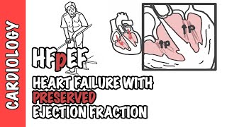
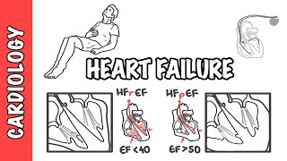
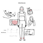
Overview
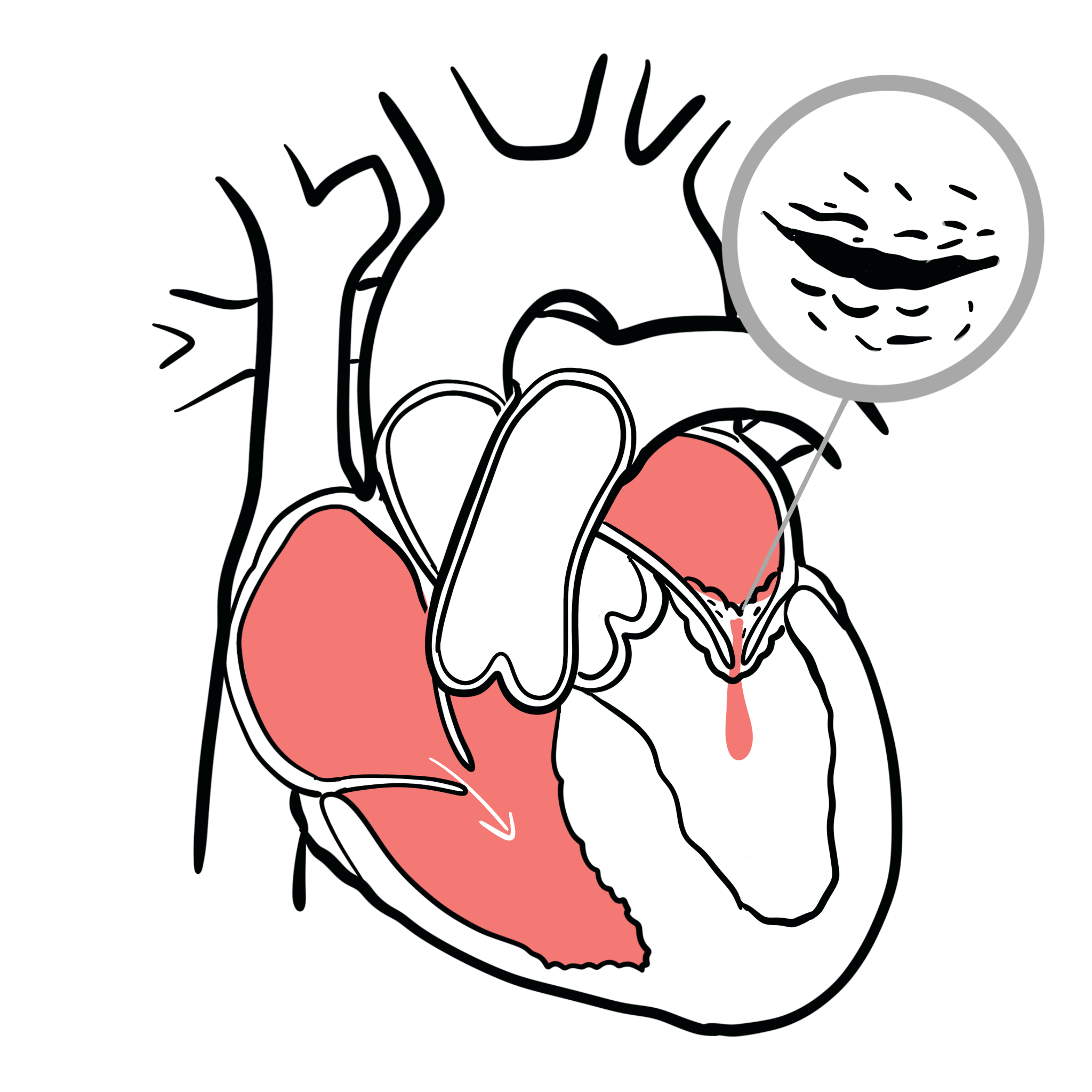
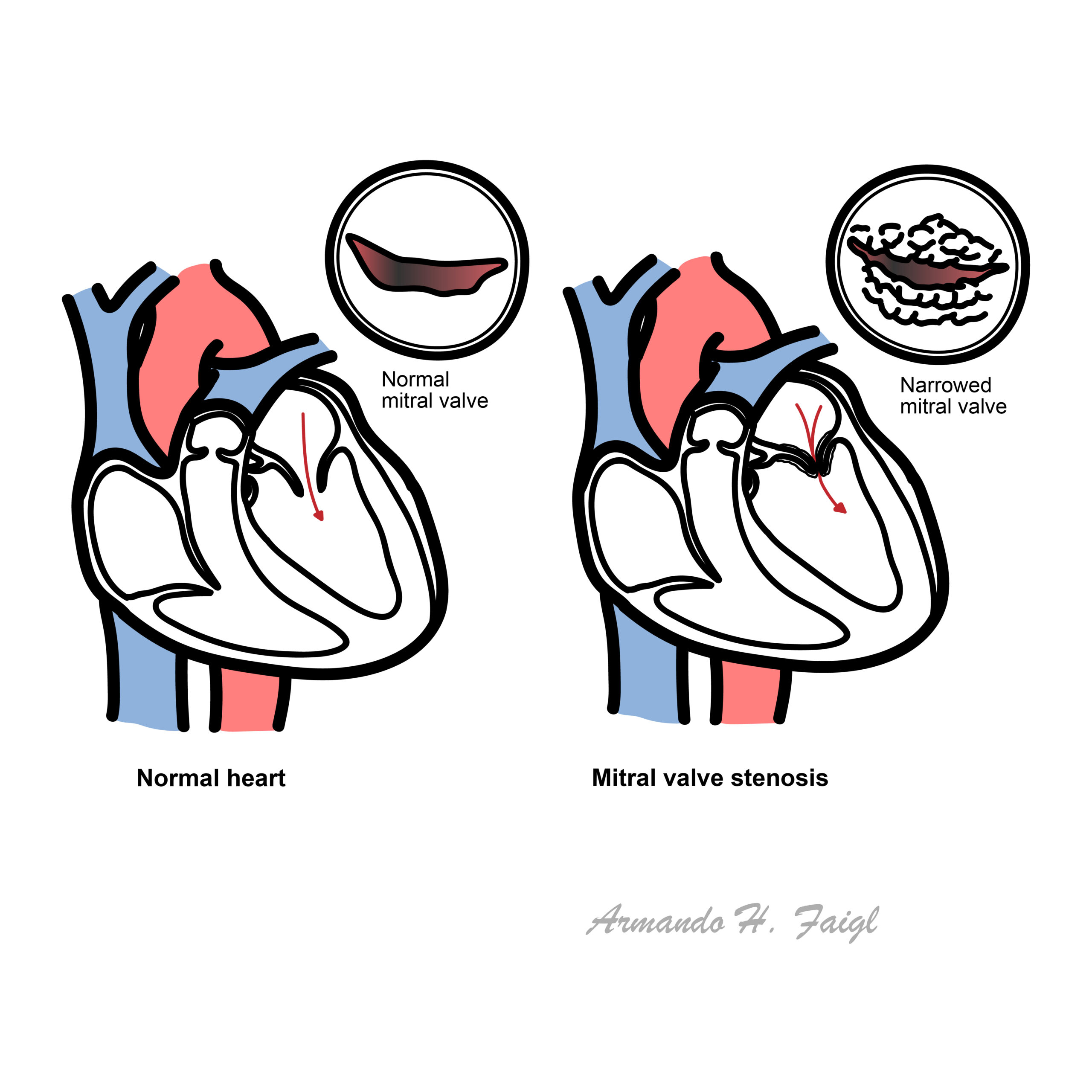
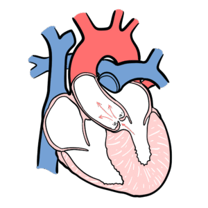
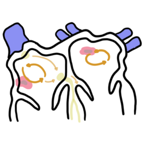
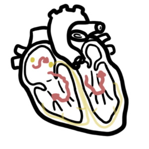
Mitral stenosis causes an obstruction to blood flow from the left atrium to left ventricle usually as a result of rheumatic heart disease. The stenosis results in increased pressure in the left atrium, pulmonary vasculature and right side of heart. Mitral valve disease is a frequent cause of heart failure and death.
Valvular Heart Disease
Endocarditis
Aortic Valve Disease
Mitral Valve Disease
Pulmonary Valve Disease
Tricuspid Valve Disease
Aetiology and Risk Factors
Aetiology
Rheumatic fever leading to rheumatic heart disease (95% of cases)
Congenital
Risk Factors
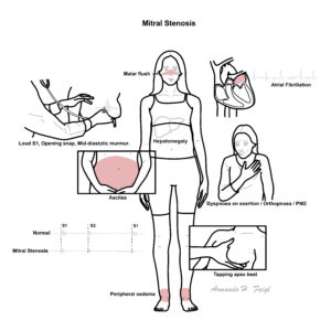
Clinical Manifestation
Exertional dyspnoea
Decreased excercise tolerance
Haemoptysis
Chest pain
Fatigue
History of rheumatic fever
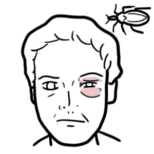
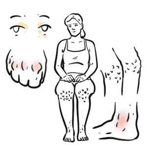
Malar flush
Signs of right-sided heart failure
Thromboembolic event “Stroke ”Hoarseness (recurrent laryngeal nerve compressed)
Dysphagia (esophagus compressed)
This image series is only available to members
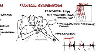
Cardiovascular Examination
Malar flush
Pulse
Left parasternal heave (from right ventricular hypertrophy)
Auscultation – Mitral valve (Apex – left 5th intercostal space mid-clavicular)
Pre systolic murmur precedes S1, a result of increase blood flow from atrial contraction
Opening snap of the mitral valve following S2 (closure of the aortic and pulmonic valves) is the opening of the stenotic mitral valve (SNAP)
Long murmur during Diastole (longer in chronic mitral stenosis)
Low-pitched diastolic rumble that is most prominent at the apex.
Early diastolic murmur (on inspiration) due to pulmonary regurgitation from pulmonary hypertension (Graham Steell murmur) may be heard rarely.
Diagnosis
Symptoms and signs similar to mitral stenosis
left atrial myxoma
prosthetic valve obstruction
Cor tratriatum
Investigations
ECG
Atrial fibrillation
Left atrial enlargement – P mitrale
Right ventricular hypertrophy – Right axis deviation
Chest X-ray
Straight or convex L heart border
Double shadow of LA behind RA
Splaying of carina
Dilated upper lobe veins
Prominent pulmonary conus
Pulmonary haemosiderosis
Trans-thoracic echocardiography
Transoesophageal echocardiography
Cardiac catherization
Diagnosis
Echocardiography — A transthoracic echocardiogram is indicated in patients with signs or symptoms of MS to establish the diagnosis, quantify the hemodynamic severity determine the etiology , and assess concomitant valve disease.
Treatment
Medication – Preload reduction
No treatment generally required if asymptomatic but monitoring is important
Diuretics and sodium
Surgery
Balloon valvotomy
moderate to severe symptomatic disease
Diuretic
Valve replacement or repair
Mechanical
Bioprosethetic
Complications and Prognosis
Complications
Atrial Fibrillation
Stroke
Warfarin-induced haemorrhage
Systemic Embolism – due to thrombus formation in the right atrium
Infective endocarditis
Functional tricuspid reguritation
Prognosis
With continuous monitoring and ppropriate treatment prognosis is excellent
Death from Mitral stenosis is oftne due to progressive right-sided heart failure and/or pulmonary edema


Discussion