

Primary Hyperaldosteronsim (Conn Syndrome)
Overview
Primary hyperaldosteronism (also known as Conn syndrome when caused by a solitary aldosterone-producing adenoma) is an endocrine disorder characterized by the autonomous, unregulated overproduction of aldosterone from the zona glomerulosa of the adrenal cortex. Under physiological conditions, aldosterone secretion is regulated by the renin-angiotensin-aldosterone system (RAAS) and serum potassium levels. In primary hyperaldosteronism, aldosterone production becomes independent of renin, causing pathologically increased sodium reabsorption and potassium/hydrogen ion excretion in the distal nephron. This physiological derangement leads to volume expansion, suppression of plasma renin activity, secondary hypertension, hypokalemia, and metabolic alkalosis.
Epidemiologically, primary hyperaldosteronism is recognized as the single most common cause of secondary hypertension, accounting for 5% to 10% of all hypertensive adults and up to 20% of patients with resistant hypertension. It most commonly presents in adults aged 30 to 60 years, with a slight female predominance in cases driven by aldosterone-producing adenomas and a male predominance in bilateral adrenal hyperplasia. Early screening and targeted management are crucial because excess aldosterone exerts direct toxic pro-inflammatory and pro-fibrotic effects on cardiovascular and renal tissue, leading to significantly higher rates of stroke, myocardial infarction, and atrial fibrillation compared to essential hypertension of equivalent blood pressure levels.
Definition
Aldosterone-to-Renin Ratio (ARR): The primary screening blood test for primary hyperaldosteronism; calculated by dividing plasma aldosterone concentration by plasma renin activity (or direct renin concentration).
Conn Syndrome: Specifically refers to primary hyperaldosteronism caused by a solitary, benign aldosterone-producing adrenal adenoma.
Aldosterone Escape Phenomenon: Spontaneous diuresis and natriuresis occurring after initial volume expansion due to increased atrial natriuretic peptide (ANP) release and pressure natriuresis, preventing overt clinical edema in primary hyperaldosteronism.
Adrenal Venous Sampling (AVS): Gold standard invasive procedure used to differentiate unilateral (adenoma) from bilateral (hyperplasia) aldosterone hypersecretion prior to considering surgical adrenalectomy.
Glucocorticoid-Remediable Aldosteronism (GRA): Rare autosomal dominant familial form (FH-I) caused by a chimeric gene mutation where aldosterone synthesis is ectopically regulated by adrenocorticotropic hormone (ACTH).
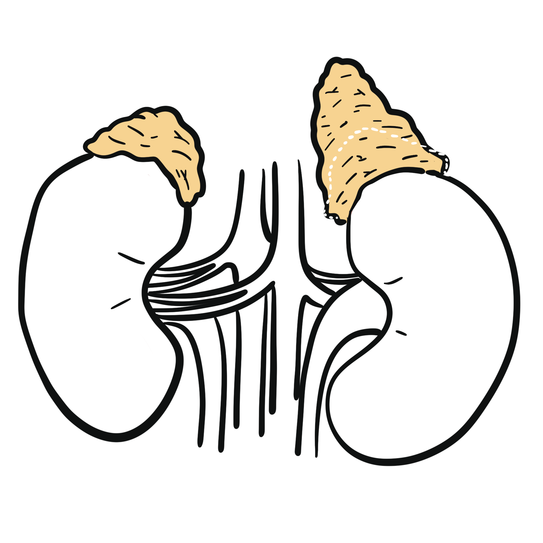
Adrenal Glands Anatomy & Physiology
Anatomy
- The normal adrenal glands weigh 4–5 g.
- The cortex represents 90% of the normal gland and surrounds the medulla.
- The arterial blood supply arises from the renal arteries, aorta, and inferior phrenic artery.
- Venous drainage occurs via the central vein into the inferior vena cava on the right, and into the left renal vein on the left.
- Three layers of the adrenal glands
- Zona fasiculata
- Zona glomerulosa
- Zona Reticularis
Physiology
- Glucocorticoids (zona fasciculata)
- Mineralcorticoids (zona glomerulosa)
- Androgens (zona reticularis and zona fasciculata)
Mineralocorticoids and aldosterone
- Glucocorticoids and mineralocorticoids are synthesised from cholesterol
Classification
Underlying Anatomical Etiology
- Bilateral Idiopathic Adrenal Hyperplasia (BAH): Accounts for ~60% of cases; characterized by diffuse or nodular hyperplastic changes in both adrenal glands. Managed medically.
- Aldosterone-Producing Adenoma (APA / Conn Syndrome): Accounts for ~35% of cases; solitary, small (< 2 cm), lipid-rich benign neoplasm of the adrenal cortex. Managed surgically.
- Primary (Unilateral) Adrenal Hyperplasia: Accounts for ~2% of cases; unilateral micro- or macronodular hyperplasia.
- Adrenocortical Carcinoma: Rare (< 1% of cases); malignant aldosterone-secreting tumor, typically large (> 4 cm) on cross-sectional imaging.
Bilateral Idiopathic Adrenal Hyperplasia (~60%) and Aldosterone-Producing Adenoma (~35%) account for over 95% of all cases of primary hyperaldosteronism.
Aetiology & Risk Factors
Aetiology
- Somatic Mutations in APA: Up to 90% of solitary aldosterone-producing adenomas carry acquired somatic driver mutations in ion channels or pumps (KCNJ5, ATP1A1, ATP2B3, CACNA1D), leading to intracellular depolarization and autonomous CYP11B2 (aldosterone synthase) transcription.
- Genetic Inherited Syndromes: Familial Hyperaldosteronism types I-IV (autosomal dominant).
Indications for Screening High-Risk Populations
- Sustained hypertension with blood pressure > 150/100 mmHg on three separate days.
- Resistant hypertension (uncontrolled BP despite adherence to three antihypertensive drugs, including a diuretic).
- Controlled BP requiring four or more antihypertensive medications.
- Hypertension accompanied by spontaneous or diuretic-induced hypokalemia.
- Hypertension with an incidentally discovered adrenal adenoma (incidentaloma).
- Hypertension with a family history of early-onset hypertension or cerebrovascular disease at an age < 40 years.
- Hypertension in all first-degree relatives of patients diagnosed with primary hyperaldosteronism.
Hypokalemia is NOT required to suspect or diagnose primary hyperaldosteronism. Over 60% of patients with primary hyperaldosteronism are normokalemic at presentation!
Pathophysiology
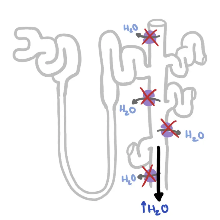
- Renal Epithelial Actions: Aldosterone binds to intracellular mineralocorticoid receptors in the distal nephron principal cells, upregulating luminal Epithelial Sodium Channels (ENaC) and basolateral Na+/K+-ATPase pumps. This increases Na+ reabsorption and creates a negative lumen potential that drives K+ secretion (via ROMK channels) and H+ secretion (via intercalated cell H+-ATPase pumps).
- Suppression of Renin: Fluid retention increases effective arterial blood volume and renal perfusion pressure, completely suppressing juxtaglomerular apparatus renin release (Plasma Renin Activity < 1.0 ng/mL/hr).
- Aldosterone Escape Mechanism: Initial sodium and water retention expands extracellular volume by 1 to 2 liters. Increased atrial stretch releases Atrial Natriuretic Peptide (ANP) and triggers pressure natriuresis, halting further sodium retention and preventing clinical edema.
- Cardiovascular & Renal Toxicity: Independent of blood pressure, excess aldosterone induces systemic vascular inflammation, endothelial dysfunction, myocardial fibrosis, arterial stiffness, and glomerular hyperfiltration.
The “Aldosterone Escape” phenomenon explains why patients with primary hyperaldosteronism present with hypertension and hypervolemia, but NEVER present with peripheral edema in the absence of co-existing heart or liver failure.
Clinical Manifestations
Cardiovascular Manifestations
- Secondary Hypertension
- Nocturnal Non-Dipping: Absence of normal physiological blood pressure drop during sleep on 24-hour ambulatory blood pressure monitoring.
- Left ventricular hypertrophy
- Stroke
- Coronary artery disease
- Heart failure
- Atrial fibrillation
Neuromuscular & Hypokalemic Manifestations
- Muscle Weakness & Cramps: Proximal muscle fatigue, nocturnal leg cramps, or transient flaccid paralysis (in severe hypokalemia, K+ < 2.5 mEq/L).
- Paresthesias & Tetany: Secondary to hypocalcemia/hypomagnesemia induced by metabolic alkalosis.
- Polyuria & Polydipsia: Impaired renal concentrating ability (nephrogenic diabetes insipidus) caused by chronic intracellular renal tubular potassium depletion.
Polyuria and polydipsia in a patient with severe hypertension are classic signs of hypokalemia-induced nephrogenic diabetes insipidus secondary to hyperaldosteronism.
Diagnosis
Diagnosis follows a strict 3-step pathway: Screening, Confirmatory Testing, and Subtype Classification (Localization).
Step 1: Initial Screening (Aldosterone-to-Renin Ratio – ARR)
- Pre-Test Preparation: Correct hypokalemia first (hypokalemia suppresses aldosterone secretion, causing false-negative results). Discontinue mineralocorticoid receptor antagonists (Spironolactone, Eplerenone) for at least 4 to 6 weeks before testing.
- Testing Conditions: Morning blood sample drawn after the patient has been ambulatory for at least 2 hours and seated for 5 to 15 minutes.
- Screening Result: Positive if ARR > 20 to 30 (when plasma aldosterone concentration is expressed in ng/dL and plasma renin activity in ng/mL/hr) AND Plasma Aldosterone Concentration (PAC) is elevated (> 15 ng/dL).
Step 2: Confirmatory Suppression Testing
Required to demonstrate autonomous, non-suppressible aldosterone production (can be skipped if patient presents with spontaneous hypokalemia, undetectable renin, and PAC > 20 ng/dL):
- Oral Sodium Loading Test: High-sodium diet for 3 days; 24-hour urinary aldosterone > 12 mcg/24 hr confirms diagnosis.
- Intravenous Saline Infusion Test: 2 liters of 0.9% normal saline infused IV over 4 hours. Post-infusion PAC > 10 ng/dL confirms primary hyperaldosteronism (PAC < 5 ng/dL rules it out).
- Fludrocortisone Suppression Test & Captopril Challenge Test: Alternative confirmatory protocols.
Step 3: Subtype Classification & Localization
- Non-Contrast Adrenal CT Scan: First-line imaging modality to visualize adrenal anatomy, identify solitary adenomas, and rule out adrenocortical carcinoma.
- Adrenal Venous Sampling (AVS – Gold Standard)
- Mandatory prior to surgery in patients > 35 years old to confirm lateralization (unilateral adenoma vs. bilateral hyperplasia)
- CT scans frequently misidentify non-functioning incidentalomas or miss microadenomas.
- Genetic Testing: Indicated for patients with onset of primary hyperaldosteronism < 20 years old or with a family history of early-onset disease/stroke (tests for CYP11B1/CYP11B2 chimeric gene in GRA).
Before measuring the Aldosterone-to-Renin Ratio (ARR), Spironolactone and Eplerenone MUST be stopped for at least 4 to 6 weeks, as they falsely elevate renin levels and cause false-negative screening results.
Primary vs Secondary Hyperaldosteronsim
| Feature | Primary Hyperaldosteronism | Secondary Hyperaldosteronism |
| Site of Primary Pathology | Adrenal cortex (zona glomerulosa) | Extra-adrenal (Renal, Cardiovascular, Hepatic) |
| Plasma Renin Activity (PRA) | Suppressed / Low (< 1.0 ng/mL/hr) | Elevated / High |
| Plasma Aldosterone (PAC) | Elevated (> 15 ng/dL) | Elevated |
| Aldosterone-to-Renin Ratio (ARR) | High (> 20–30) | Normal or Low (< 10–15) |
| Most Common Etiologies | Bilateral Adrenal Hyperplasia (~60%), Aldosterone-Producing Adenoma (~35%) | Renal Artery Stenosis, Heart Failure, Cirrhosis, Nephrotic Syndrome, Diuretic Therapy |
| Blood Pressure | Secondary Hypertension (often severe or resistant) | Variable (Hypertension in RAS; Normal or Low in Cirrhosis/Heart Failure) |
| Peripheral Edema | Absent (due to “Aldosterone Escape”) | Often Present in HF/Cirrhosis/Nephrotic syndrome; Absent in RAS |
| Serum Potassium | Hypokalemia (in ~40% of cases) with Metabolic Alkalosis | Hypokalemia (frequent, especially with diuretic co-use) |
| Diagnostic Workup | Screening ARR -> Confirmatory Saline Suppression Test -> Adrenal CT / AVS | Renal Duplex US / CT Angiography, Echocardiogram, Hepatic Workup |
| Primary Management | Laparoscopic Adrenalectomy (Unilateral) or Spironolactone/Eplerenone (Bilateral) | Treat underlying cause (e.g., Revascularization for RAS, Medical optimization for HF/Cirrhosis) |
Treatment
Treatment strategy depends entirely on whether disease is unilateral (surgical candidate) or bilateral (medical management).
Unilateral Disease (Aldosterone-Producing Adenoma / Unilateral Hyperplasia)
- Surgical Treatment (Gold Standard):Laparoscopic Adrenalectomy of the affected gland.
- Pre-Operative Preparation: Treat with Spironolactone or Eplerenone for 4 to 8 weeks to normalize blood pressure and potassium levels prior to surgery.
- Outcomes: Cures hypokalemia in 100% of cases and improves or cures hypertension in 50% to 80% of patients.
Bilateral Disease (Bilateral Idiopathic Adrenal Hyperplasia / Non-Surgical Candidates)
- Medical Treatment (First-Line):Mineralocorticoid Receptor Antagonists (MRAs).
- Spironolactone: Competitive MRA; highly effective first-line drug (starting dose 12.5–25 mg daily). Side effects: dose-dependent gynecomastia, mastodynia, erectile dysfunction, and menstrual irregularities due to progesterone and androgen receptor binding.
- Eplerenone: Selective MRA; lacks anti-androgenic side effects and is preferred if spironolactone causes intolerable gynecomastia or breast tenderness.
- Adjunctive Therapy: Amiloride or Triamterene (ENaC blockers) if MRAs are contraindicated; thiazide or loop diuretics to assist volume control.
Glucocorticoid-Remediable Aldosteronism (GRA / FH-I)
- Medical Treatment: Low-dose synthetic glucocorticoid (Dexamethasone or Prednisone) to suppress endogenous pituitary ACTH secretion and halt chimeric aldosterone production.
Laparoscopic Adrenalectomy is the definitive treatment of choice for unilateral Aldosterone-Producing Adenoma (Conn Syndrome), whereas Spironolactone or Eplerenone is the first-line treatment for Bilateral Adrenal Hyperplasia.
Complications & Prognosis
- Cardiovascular Morbidity
- Left ventricular hypertrophy
- Myocardial infarction
- Stroke, transient ischemic attack
- Atrial fibrillation
- Renal Dysfunction
- Glomerular hyperfiltration initially masks underlying renal damage; initiating MRA therapy or performing adrenalectomy leads to an unmasking drop in eGFR.
- Metabolic Abnormalities
- Increased incidence of metabolic syndrome
- Type 2 Diabetes Mellitus (aldosterone impairs pancreatic beta-cell insulin secretion and reduces peripheral insulin sensitivity).
- Excellent; surgical resection or targeted MRA therapy normalizes serum potassium, controls blood pressure, reverses left ventricular hypertrophy, and reduces cardiovascular event rates back toward baseline levels.
- Persistent exposure to excess aldosterone leads to irreversible systemic vascular fibrosis, progressive chronic kidney disease, and permanent essential-like vascular resistance.
A drop in eGFR after starting Spironolactone or after unilateral adrenalectomy is expected and reflects the resolution of aldosterone-induced glomerular hyperfiltration, rather than drug toxicity or surgical injury.
References
- Funder JW, Carey RM, Mantero F, Murad MH, Reincke M, Shibata H, et al. The Management of Primary Aldosteronism: Case Detection, Diagnosis, and Treatment: An Endocrine Society Clinical Practice Guideline. J Clin Endocrinol Metab. 2016;101(5):1889-1916. doi:10.1210/jc.2015-4061
- Vaidya A, Mulatero P, Baudrand R, Adler GK. The Changing Definition of Primary Aldosteronism. J Am Coll Cardiol. 2018;71(20):2362-2372. doi:10.1016/j.jacc.2018.03.460
- Young WF Jr. Diagnosis and treatment of primary aldosteronism: practical clinical perspectives. J Intern Med. 2019;285(2):126-148. doi:10.1111/joim.12831


Members only discussions coming soon…