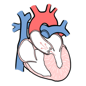
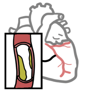
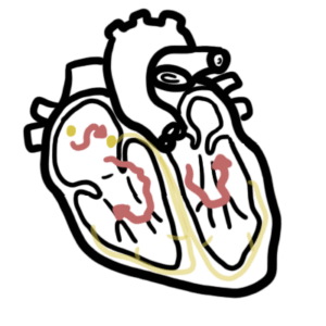
0:00 In this video we will look at cardiovascular diseases and we'll break them down 0:10 into categories. 0:12 So the first and the most common is the coronary artery disease, also known as 0:20 ischemic heart 0:21 disease. 0:22 And it is where we have poor blood flow to the heart. 0:26 And here we have the heart, and the heart is supplied by the blood, by vessels 0:32 called 0:32 the coronary arteries. 0:34 But if we have a plaque as a result of atherosclerosis for example, we have 0:39 decreased blood flow 0:41 to the heart tissues. 0:43 And if we get no blood flow, it can lead to acute myocardial infarction. 0:49 And this is where we have necrosis of the myocardial tissue caused by lack of 0:55 oxygenation 0:56 and blood flow. 0:58 And as I mentioned, acute myocardial infarction is a result of an occluded 1:05 coronary artery. 1:06 So coronary artery disease is associated with angina, which occurs when there 1:12 is a temporary 1:14 loss of blood supply to the heart. 1:16 And this causes chest pain. 1:19 There are two types of angina, stable angina and unstable angina. 1:24 Stable angina is where we have a stable plaque, and chest pain comes about upon 1:30 exertion, 1:30 so exercising. 1:33 Then there is unstable angina, which is a result of a hemodynamically unstable 1:39 plaque. 1:40 And we have thrombosis involved. 1:43 A heart attack occurs when the heart blood vessel is suddenly blocked. 1:48 And this is life-threatening and leads to myocardial infarction, as mentioned. 1:56 The next type of cardiovascular disease we will look at is heart failure. 2:00 Now heart failure can result from many heart and body conditions, disorders 2:06 such as coronary 2:07 heart disease, hypertension, cardiomyopathies, and vasculitis, amongst many 2:13 other things. 2:15 Heart failure is where the heart is basically unable to maintain a strong blood 2:20 flow and 2:20 pump it to the body around the body. 2:23 And this results in chronic tiredness, reduced physical activity, and shortness 2:29 of breath. 2:30 So here we have the heart, and it's for chambers, and here we have the inferior 2:35 vena cava, which 2:36 connects with the liver via the hepatic vein, and the liver coming out of the 2:41 liver from 2:42 the bottom, we have the portal vein. 2:49 Heart failure can be divided into three types. 2:51 Right-sided heart failure, left-sided heart failure, and congestive heart 2:57 failure. 2:57 Right-sided heart failure is usually a result of left-sided heart failure. 3:02 In right-sided heart failure, so we're talking about the right atrium and right 3:08 ventricle, 3:09 blood can back up into other tissues, such as the liver and the abdomen, 3:15 causing congestion 3:17 in these areas. 3:20 So as a result of right-sided heart failure, we can have hepatomegaly and asc 3:27 ites because 3:28 of the back flow from the right ventricle and right atrium, because the right 3:33 side of 3:34 the heart fails to pump blood out, to pump blood in the right direction. 3:40 Now in left-sided heart failure, oxygenated blood, because on the left side we 3:45 have the 3:45 left ventricle and left atrium, which contains the oxygenated blood from the 3:50 lungs. 3:51 So in left-sided heart failure, oxygenated blood cannot be pumped out from the 3:55 heart 3:56 to the rest of the body. 3:58 And so blood can back flow and accumulate in the lungs of the veins, pulmonary 4:04 veins 4:05 essentially causing fluid accumulation in the lungs, leading to shortness of 4:09 breath 4:09 and pulmonary edema. 4:14 The third type of heart failure is congestive heart failure, which involves 4:17 both right and 4:18 left-sided heart failures, leading to congestion in the lungs, pulmonary edema, 4:23 and congestion 4:24 of the liver and abdominal area. 4:27 We have a decreased cardiac output, like in all heart failures, which leads to 4:31 decreased 4:32 in venous return to the heart, which results in decreased in stroke volume and 4:36 cardiac 4:36 output again. 4:38 The cycle continues if the underlying problem is not solved. 4:44 The next cardiovascular disease is cardiomyopathies. 4:49 Now cardiomyopathies involve the muscles of the heart, hence cardio-hot myomus 4:54 selpathies 4:54 abnormality. 4:57 So cardiomyopathies is where the heart muscles become enlarged, thickened, or 5:05 stiff, reducing 5:06 the effectiveness of the heart, leading to heart failure. 5:12 There are three main types of cardiomyopathies, dilated cardiomyopathy, hypert 5:17 rophic cardiomyopathy, 5:19 and restrictive cardiomyopathy. 5:22 Let's look at dilated first. 5:25 Dilated cardiomyopathy is when there is less blood pumped from the heart, 5:30 because ventricles 5:31 are enlarged and weakened, and the dilated cardiomyopathy can lead to systolic 5:38 heart failure with 5:40 a big decrease in ejection fraction. 5:47 Here we have enlarged ventricles with, yeah, dilation. 5:54 Hypertrophic cardiomyopathy, on the other hand, is when there's less blood 6:00 pumped from 6:00 the heart, because the ventricles can't fully relax. 6:04 So it's a problem in diastole, and thus, hypertrophic cardiomyopathy can lead 6:09 to diastolic heart failure. 6:12 So hypertrophic cardiomyopathy, we have a thick ventricle, super thick, and the 6:19 muscles of 6:20 the ventricles are super thick, and the ventricular septum is also thick, thus 6:24 restricting proper 6:25 feeling of the ventricles with blood. 6:30 Restrictive cardiomyopathy is when the left ventricle maintains normal 6:35 dimensions, but 6:37 we develop left atrial hypertrophy and dilation, and right ventricular hypertro 6:43 phy as a result. 6:44 So here we have hypertrophy and dilation of the left atria, and hypertrophy of 6:50 the right 6:51 ventricle as a result, because of the backflow from the left side of the heart 6:59 to the right. 7:01 The next cardiovascular disease involves the aorta, so we are going to classify 7:06 this as 7:06 aortic disease. 7:08 An example is when we have an abnormal widening of the aorta as an aortic aneur 7:14 ysm, specifically 7:16 abdominal aortic aneurysm. 7:19 So if we look at this aortic aneurysm here, it results in a weakened aortic 7:24 wall and 7:25 plaque formation, and this aortic aneurysm can result in thrombosis and then 7:32 embolist, 7:33 which can, you know, lodge into vessels leading to some very bad problems 7:41 downstream. 7:43 Another aortic disease is aortic dissection, when blood will fill the walls of 7:49 the aorta 7:50 vessel. 7:52 So this is due to some disruption or abnormality in the aortic vessel wall, and 7:58 allowing blood 7:59 to accumulate between the tunical layers of the aorta. 8:10 Peripheral vascular disease, also known as peripheral arterial disease, is 8:15 another cardiovascular 8:17 disease. 8:19 So here we have the heart with a descending aorta. 8:23 Peripheral vascular disease is basically a result of plaque formation in the 8:27 peripheral 8:28 vasculature from atherosclerosis, for example. 8:37 So obstruction of large arteries that supply blood to the peripheries, and this 8:43 can lead 8:44 to devastating like big problems, such as if we have a renal stenosis, for 8:52 example. 8:53 Another cardiovascular disease is a category is a valvulitis, or more 8:59 specifically inflammation 9:01 of the valves, or more specifically, valvular disease of the heart. 9:09 So inflammation of the valve, the most common cause, is rheumatic heart disease 9:14 . 9:14 Let's focus on rheumatic heart disease. 9:17 Rheumatic heart disease is where there is permanent damage to the heart muscle, 9:20 mainly the valves 9:22 caused by rheumatic fever. 9:25 So if we look at rheumatic fever and how it comes about, well, a bacteria 9:31 called group 9:32 A strep, which are basically your streptococcus pyogenes, can cause pharyngitis 9:39 in the throat, 9:41 which then can cause rheumatic fever, which then can lead to rheumatic heart 9:45 disease. 9:46 Or group A streptococcus can lead straight to rheumatic fever as a result of 9:53 local skin 9:54 infection, for example. 9:56 Other valvular disease, which do not always involve inflammation of the valves, 10:02 include 10:02 aortic stenosis, aortic regurgitation, mitral stenosis, and mitral regurg 10:10 itation. 10:12 The next cardiovascular disease is pericarditis, which is inflammation of the 10:17 pericardium. 10:18 The pericardium is a fibrous and serous layer protecting the heart. 10:23 We will look at how the pericardium looks like inflamed and how it looks like, 10:29 normally, 10:30 using a diagrammatic representation. 10:33 So when the pericardium is normal, this is what it looks like. 10:37 You have the outer fibrous pericardium, the parietal layer, the parietal layer, 10:43 and then 10:44 we have the visceral pericardium, which directly surrounds the heart. 10:49 In between the parietal and visceral layer of the pericardium is the pericardio 10:54 cavity, 10:55 which is a cavity that contains pericardio fluid that serves to reduce friction 11:01 between 11:01 the pericardium. 11:04 In an inflamed pericardium, we have disruption of the pericardio layers, 11:10 inflammation, with 11:11 thickening and damage to the pericardium, resulting in friction of the pericard 11:17 io layers 11:18 and pain. 11:19 So the pericardium rub against each other as the heart contracts and relaxes. 11:27 Other pericardio diseases are pericardio effusion, which can lead to tamponade 11:34 and hemopericardium, 11:36 which is life-threatening. 11:41 The last cardiovascular disease we will look at is the congenital heart 11:45 diseases, which 11:46 are disorders of the heart or the central blood vessels that is present at 11:52 birth. 11:53 The congenital heart diseases are actually the leading cause of death in the 11:58 first year 11:59 of life. 12:01 We will look at five main ones. 12:06 The furamin ovali is where the furamin ovali does not close, resulting in a 12:11 left to right 12:12 shunt. 12:14 Patent ductus arteriosus is where the ductus arteriosus does not close, 12:20 resulting in a 12:21 left to right shunt as well, between the aorta and pulmonary artery. 12:28 One of the aorta is another congenital heart disease. 12:36 Transposition of the great vessels is where both the furamin ovali and patent 12:41 ductus do 12:41 not close. 12:43 And the final congenital heart disease we will mention is tetralogy of fallow, 12:50 which is composed, 12:52 which is composed of four tetra things. 12:58 These are the right ventricular hypertrophy, the ventricular septal defect, 13:03 pulmonary stenosis, 13:06 and the overriding aorta. 13:07 So all these four represent tetralogy of fallow. 13:12 I hope you enjoyed this video on the main cardiovascular diseases. 13:18 Hopefully there will be some links that will go into more detail into each of 13:21 these conditions. 13:23 Thank you very much for watching. 13:24 Hope you enjoyed it. 13:28 [BLANK_AUDIO]