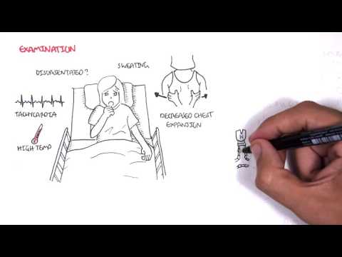
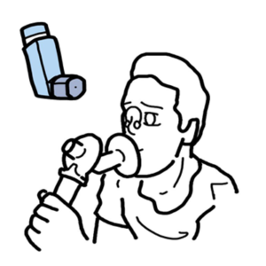
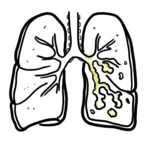
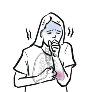
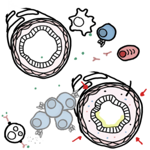
0:00 So in this video we're going to look at atelectasis which is the collapsed, 0:12 which essentially means 0:13 collapsed lung tissue. 0:16 So here is a person with a healthy lung on the right side and a collapsed lung 0:21 on the 0:22 left. 0:24 This is the lung in red. So running a lung, the lung is a layer known as the 0:30 visceral 0:31 pleura and the second layer is the parietal pleura. 0:37 If we look into the most distal aspect of the respiratory tract, we can find 0:43 the alveoli 0:44 where gas exchange takes place. 0:47 Before focusing on the collapsed lung, let us look at what keeps the lung 0:52 inflated and 0:52 what prevents it from collapsing. 0:56 So a few things does this. One, continuous breathing keeps the alveoli open. 1:02 Oxygen comes 1:03 in and carbon dioxide goes out. 1:06 Number two, coughing. Coughing clears up the air away from problems that can 1:09 lead to a 1:10 collapsed lung. 1:13 Three, cells such as type two pneumocytes secrete surfactant which prevents the 1:21 lungs from 1:22 collapsing. It keeps the alveoli basically inflated and big. 1:28 Four, normal VQ, normal gas exchange between the capillary and the alveoli 1:33 keeps the lung 1:34 healthy and prevents collapse. 1:36 Okay, so those are the things that keeps the lungs inflated and prevents it 1:41 from being 1:41 collapsed. So atelectasis means collapsed lung and there are many, many causes. 1:49 Collapse in general, compression, obstruction and surgery. 1:54 Let us begin with the general collapse. So here we have a collapsed lung. Now, 1:59 a collapsed 2:00 lung is where air can fill the plural cavity and this can be from an external 2:07 trauma such 2:08 as a puncture of the parietal pleura or it can be from an internal trauma such 2:16 as puncture 2:17 through the alveoli, the visceral pleura. Either way, the result is gas moving 2:23 into the plural 2:24 space, the gap between the parietal and visceral pleura. Thus, we have air 2:31 moving into the space 2:32 which will lead to collapsed lung. Examples of this type of atelectasis is 2:40 pneumothorax. 2:42 One can also lead to atelectasis. This can be when fluid accumulates in the 2:47 plural space 2:48 which compresses the lung tissue. Examples of this is hydrothorax. 2:58 Obstruction such as a tumor or lymphid anopathy can lead to atelectasis. 3:05 Finally, surgery can 3:06 also lead to atelectasis. For example, use of anesthesia and opioids 3:10 decreases the cough reflex and decreases deep breathing which means that air 3:15 cannot enter 3:16 the lungs properly and this can decrease lung volume which could lead to lung 3:21 collapse. 3:22 Also, because the cough reflex is decreased, aspiration can occur which can 3:29 subsequently 3:30 lead to lung problems. Now, let us see what you can, what you find upon 3:37 examination with 3:38 someone who has a collapsed lung. Upon examination, you can hear decreased 3:44 breath sounds in the 3:45 region affected when you oscillate. It is dull on percussion. Lung expansion on 3:54 affected 3:54 side decreases. Next, the radiological findings, imaging, is actually used for 4:02 diagnosis. They 4:04 definitely have it if you use imaging. What you find on imaging, you can see on 4:10 X-ray, 4:11 for example, an increase in density, opacity on the atelectic portion of the 4:17 lung. There 4:18 is displacement of fissures towards area of atelectasis. It can include 4:25 compensation where 4:26 you get overinflation of the unaffected side and it may or may not include 4:32 displacement 4:33 of thoracic structures. Displacement, if there is displacement, you would see 4:38 in cases of 4:40 attention pneumothorax. Treatment for atelectasis include maximizing coughing, 4:47 deep breathing 4:48 and releasing the tension if it is in the plural space through puncturing of 4:53 the parietal 4:54 plural. So, I hope you enjoyed this video on atelectasis. Thank you for 5:00 watching. Bye. 5:03 You