

ANCA – Associated Vasculitis with Pulmonary Haemorrhage or Fibrosis

Overview
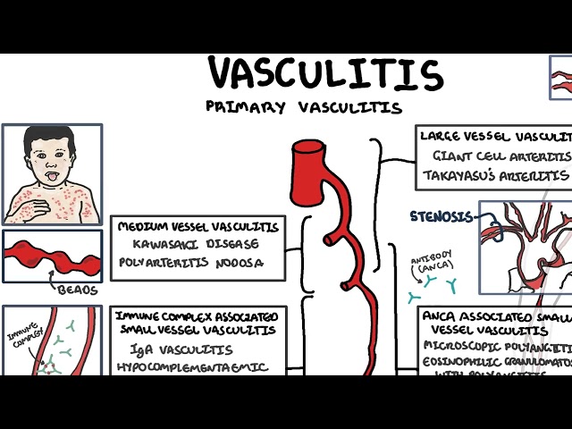
Antineutrophil cytoplasmic antibody-associated vasculitis (AAV) is a group of necrotising small-vessel vasculitides comprising granulomatosis with polyangiitis (GPA), microscopic polyangiitis (MPA) and eosinophilic granulomatosis with polyangiitis (EGPA). Pulmonary involvement ranges from acute, life-threatening diffuse alveolar haemorrhage to chronic interstitial lung disease and pulmonary fibrosis.1,2
Diffuse alveolar haemorrhage usually reflects pulmonary capillaritis and may occur as part of a pulmonary–renal syndrome. Pulmonary fibrosis is most strongly associated with myeloperoxidase-ANCA-positive MPA and may precede, accompany or follow the diagnosis of systemic vasculitis.3
Definition
- ANCA
- Autoantibodies directed against neutrophil cytoplasmic antigens, most commonly proteinase 3 or myeloperoxidase.
- Diffuse alveolar haemorrhage
- Bleeding into the pulmonary alveoli caused by disruption of the alveolar microcirculation.
- Pulmonary capillaritis
- Neutrophil-mediated inflammation and destruction of alveolar capillary walls, often producing diffuse alveolar haemorrhage.
- Pulmonary–renal syndrome
- The combination of diffuse alveolar haemorrhage and rapidly progressive glomerulonephritis.
- AAV-associated interstitial lung disease
- Inflammatory or fibrotic disease of the lung interstitium associated with ANCA positivity or systemic ANCA-associated vasculitis.
Classification
ANCA-Associated Vasculitis
- Granulomatosis with polyangiitis: commonly associated with proteinase 3-ANCA and involving the upper airways, lungs and kidneys.
- Microscopic polyangiitis: commonly associated with myeloperoxidase-ANCA; pulmonary capillaritis, glomerulonephritis and fibrotic interstitial lung disease are characteristic.
- Eosinophilic granulomatosis with polyangiitis: occurs with asthma and eosinophilia; pulmonary infiltrates are common, although alveolar haemorrhage and pulmonary fibrosis are less characteristic.
ANCA specificity is clinically informative but does not define the diagnosis by itself. Proteinase 3 and myeloperoxidase antibodies can occur outside primary vasculitis, while some patients with biopsy-proven AAV are ANCA-negative.
Pulmonary Phenotype
- Diffuse alveolar haemorrhage caused by pulmonary capillaritis.
- Pulmonary nodules, masses or cavitation, particularly in GPA.
- Tracheobronchial or subglottic disease.
- Interstitial lung disease, frequently with a usual interstitial pneumonia pattern.
- Progressive pulmonary fibrosis.
- Pleuritis or pleural effusion, less commonly.
Aetiology & Risk Factors
Aetiology
AAV develops through an interaction between genetic susceptibility, loss of immune tolerance and environmental exposures. Pro-inflammatory signals prime neutrophils, causing proteinase 3 or myeloperoxidase antigens to appear on the cell surface. ANCA binding activates neutrophils, which adhere to vascular endothelium and release reactive oxygen species, proteases and neutrophil extracellular traps. Complement activation, particularly through the alternative pathway and C5a, amplifies vascular injury.1
In pulmonary capillaritis, inflammation disrupts the alveolar capillary basement membrane, allowing erythrocytes to enter the alveoli. Recurrent subclinical or overt alveolar injury may contribute to fibrosis, although the relationship between AAV and interstitial lung disease is multifactorial. Fibrotic abnormalities can precede detectable systemic vasculitis.
Risk Factors
- MPO-ANCA or MPA: strongly associated with interstitial lung disease and pulmonary fibrosis.
- PR3-ANCA or GPA: associated with pulmonary nodules, cavitation and granulomatous airway disease.
- Previous diffuse alveolar haemorrhage.
- Older age, smoking and reduced pulmonary reserve.
- Environmental silica exposure.
- Concurrent renal involvement and pulmonary–renal disease.
- Immunosuppression, which increases infection risk and may mimic active vasculitis.
Pathophysiology
Diffuse Alveolar Haemorrhage
Activated neutrophils injure the pulmonary capillary endothelium, producing necrotising capillaritis. Blood enters the alveolar spaces and interferes with gas exchange, causing ventilation–perfusion mismatch, diffusion impairment and hypoxaemia. Haemoglobin may fall rapidly, although the initial measurement can remain normal before haemodilution occurs.
Macrophages ingest intra-alveolar erythrocytes and convert haemoglobin to haemosiderin. Haemosiderin-laden macrophages require time to develop and therefore support subacute or recurrent haemorrhage more strongly than very recent bleeding.
Interstitial Lung Disease and Fibrosis
Repeated epithelial and microvascular injury activates fibroblasts and promotes extracellular matrix deposition. MPO-ANCA-associated disease most often produces a usual interstitial pneumonia pattern, characterised by basal and subpleural fibrosis, traction bronchiectasis and honeycombing. Other patterns include nonspecific interstitial pneumonia and organising pneumonia.3
It remains uncertain whether ANCA directly drives fibrosis in every patient. Some individuals initially present with apparently idiopathic interstitial lung disease and later develop MPO-ANCA positivity or systemic MPA.
In an older patient with fibrotic interstitial lung disease, constitutional symptoms, haematuria, proteinuria, purpura, neuropathy or an unexplained inflammatory response should prompt assessment for MPO-ANCA-associated vasculitis.
Clinical Manifestations
Diffuse Alveolar Haemorrhage
The classical triad is haemoptysis, falling haemoglobin and new bilateral pulmonary infiltrates. The complete triad is frequently absent, and haemoptysis may be absent despite substantial alveolar bleeding.
- Rapidly progressive dyspnoea, cough and tachypnoea.
- Hypoxaemia and increased work of breathing.
- Diffuse crackles.
- Acute respiratory failure.
- Fever or systemic inflammatory symptoms.
Concurrent glomerulonephritis may cause haematuria, proteinuria, red-cell casts, hypertension, oedema or acute kidney injury. Other systemic manifestations include chronic sinus disease, otitis, nasal crusting, purpura, mononeuritis multiplex, inflammatory eye disease and constitutional symptoms.
The absence of haemoptysis does not exclude diffuse alveolar haemorrhage. Suspect it when unexplained hypoxaemia, falling haemoglobin and new bilateral ground-glass or consolidative opacities occur in a patient with possible systemic vasculitis.
Interstitial Lung Disease and Pulmonary Fibrosis
- Progressive exertional dyspnoea.
- Persistent dry cough and reduced exercise tolerance.
- Fine bibasal inspiratory crackles.
- Digital clubbing in advanced disease.
- Resting or exertional hypoxaemia.
- Features of pulmonary hypertension in advanced fibrosis.
Acute deterioration may reflect an exacerbation of interstitial lung disease, infection, pulmonary embolism, pulmonary oedema or diffuse alveolar haemorrhage. These possibilities require active differentiation because their treatments differ substantially.
Diagnosis & Investigations
Initial Assessment
Pulmonary haemorrhage is a medical emergency. Investigations must not unnecessarily delay treatment when life-threatening pulmonary capillaritis is strongly suspected.
Laboratory Investigations
- FBC, blood film and haemolysis studies when indicated.
- CRP and ESR
- EUC
- LFTs and Coags
- Urinalysis, urine microscopy and urine protein quantification.
- Blood cultures and targeted microbiology before immunosuppression when feasible.
- Anti-glomerular basement membrane antibodies.
- Antinuclear antibodies and complement when alternative immune-mediated disease is possible.
Test for both proteinase 3-ANCA and myeloperoxidase-ANCA using antigen-specific immunoassays. A positive result supports the diagnosis in an appropriate clinical setting but does not replace clinical assessment or tissue confirmation. ANCA titres alone should not determine whether disease is active or dictate treatment changes.2
Imaging
| Clinical setting | Typical high-resolution CT findings |
|---|---|
| Diffuse alveolar haemorrhage | Bilateral ground-glass opacity, consolidation and centrilobular opacities, with rapid evolution or clearing as haemorrhage resolves. |
| Fibrotic AAV-associated interstitial lung disease | Basal and subpleural reticulation, traction bronchiectasis, honeycombing, architectural distortion and volume loss. |
| Other AAV lung disease | Nodules, masses, cavitation, airway stenosis, pleural disease or organising pneumonia. |
Bronchoscopy
Bronchoscopy with bronchoalveolar lavage is useful when the diagnosis remains uncertain or infection must be excluded. Diffuse alveolar haemorrhage is supported by lavage aliquots that become progressively bloodier or fail to clear. Cytology may later demonstrate haemosiderin-laden macrophages. Bronchoscopy may be unsafe in severe hypoxaemia or haemodynamic instability and should not delay necessary treatment.
Histopathology
- Kidney biopsy commonly demonstrates pauci-immune necrotising and crescentic glomerulonephritis.
- Skin biopsy may demonstrate leukocytoclastic vasculitis.
- Lung biopsy may show capillaritis, granulomatous inflammation or a fibrotic interstitial pattern but is rarely required during acute haemorrhage.
Pulmonary Function and Cardiac Assessment
- Spirometry and lung volumes.
- Diffusing capacity for carbon monoxide.
- Resting and exertional oxygen saturation.
- Six-minute walk testing where appropriate.
- Serial high-resolution CT and pulmonary function testing.
- Echocardiography when pulmonary hypertension is suspected.
Important mimics of diffuse alveolar haemorrhage include infection, cardiogenic pulmonary oedema, acute respiratory distress syndrome, anticoagulant-related bleeding, anti-glomerular basement membrane disease, systemic lupus erythematosus, drug-induced vasculitis and other pulmonary capillaritis syndromes.
Treatment
Immediate Supportive Management
- Provide supplemental oxygen and ventilatory support as required.
- Correct haemodynamic instability and clinically significant anaemia.
- Stop anticoagulants when appropriate and reverse excessive anticoagulation.
- Obtain microbiological samples and commence empirical antimicrobial therapy when infection cannot be safely excluded.
- Use lung-protective mechanical ventilation for acute respiratory failure.
- Provide venous thromboembolism prophylaxis once bleeding risk permits.
Remission Induction for Organ- or Life-Threatening AAV
Diffuse alveolar haemorrhage represents organ- or life-threatening disease. Standard induction treatment combines glucocorticoids with either rituximab or cyclophosphamide. Rituximab is often preferred in relapsing or PR3-ANCA disease, patients wishing to preserve fertility, or those with substantial previous cyclophosphamide exposure. Cyclophosphamide remains appropriate for severe presentations.2,5
Intravenous methylprednisolone pulses are frequently used for immediately life-threatening pulmonary haemorrhage, followed by oral glucocorticoids. Contemporary guidelines favour reduced cumulative glucocorticoid exposure because reduced-dose regimens achieve similar major outcomes with fewer serious infections.2,5
Avacopan can reduce glucocorticoid exposure in selected GPA or MPA patients. Evidence is limited in the most severe diffuse alveolar haemorrhage requiring mechanical ventilation, and its use should not delay established emergency induction therapy.
Plasma Exchange
PEXIVAS found that routine plasma exchange did not reduce the overall risk of death or end-stage kidney disease in severe AAV. It should therefore not be added automatically for every episode of alveolar haemorrhage.6
- Consider selectively for diffuse alveolar haemorrhage with hypoxaemia.
- Consider when concurrent severe or rapidly progressive kidney failure is present.
- Use for disease overlapping with anti-glomerular basement membrane antibodies.
- Balance potential benefit against infection, bleeding and vascular-access risks.
Plasma exchange is not routine treatment for all ANCA-associated pulmonary haemorrhage. Consider it selectively—particularly for hypoxaemic haemorrhage, severe rapidly progressive renal failure or anti-GBM overlap—after multidisciplinary assessment.
Maintenance of Remission
After remission, rituximab is generally preferred for maintenance in GPA or MPA. Alternatives include azathioprine; methotrexate may be considered when kidney function is adequate and pulmonary disease is controlled. Maintenance commonly continues for at least 18–24 months and often longer when relapse risk is high. Persistent ANCA positivity or a rising titre does not, by itself, prove relapse.2
Infection Prevention and Monitoring
- Pneumocystis jirovecii pneumonia prophylaxis unless contraindicated.
- Vaccination review and infection screening.
- Monitoring of blood count, renal function and liver function.
- Immunoglobulin measurement with repeated rituximab.
- Fertility and malignancy-risk counselling before cyclophosphamide.
- Bone, gastric and metabolic protection appropriate to glucocorticoid exposure.
Interstitial Lung Disease and Pulmonary Fibrosis
- Treat active organ-threatening vasculitis with rituximab or cyclophosphamide plus glucocorticoids.
- Manage inflammatory interstitial disease through a multidisciplinary vasculitis and interstitial-lung-disease service.
- Consider antifibrotic treatment such as nintedanib when criteria for progressive pulmonary fibrosis are met; evidence is largely extrapolated from broader fibrosing ILD populations.
- Provide smoking cessation, pulmonary rehabilitation, vaccination and supplemental oxygen when indicated.
- Assess reflux, sleep-disordered breathing, pulmonary hypertension and infection.
- Consider early lung-transplant referral in appropriate patients with progressive advanced disease.
Established honeycomb fibrosis is unlikely to reverse with immunosuppression. Excessive immunosuppression in fibrosis without active vasculitis can increase harm without improving lung function.
Complications & Prognosis
Complications
- Acute hypoxaemic respiratory failure or acute respiratory distress syndrome.
- Severe anaemia and haemodynamic compromise.
- Rapidly progressive glomerulonephritis and kidney failure.
- Superimposed bacterial, viral, fungal or opportunistic infection.
- Venous thromboembolism.
- Recurrent pulmonary haemorrhage.
- Progressive pulmonary fibrosis, pulmonary hypertension and right-heart failure.
- Treatment-related cytopenia, infertility, malignancy, hypogammaglobulinaemia or glucocorticoid toxicity.
- Relapse following initial remission.
Prognosis
Diffuse alveolar haemorrhage is associated with substantial early mortality, particularly when mechanical ventilation, severe infection or advanced renal impairment is present. Prompt recognition and remission-induction therapy improve survival, but relapse and treatment toxicity remain important long-term risks.
AAV-associated interstitial lung disease confers a worse prognosis than AAV without fibrosis. Adverse prognostic features include advanced age, a usual interstitial pneumonia pattern, declining forced vital capacity, reduced diffusing capacity, exertional hypoxaemia and pulmonary hypertension.3
Long-term follow-up should monitor disease activity, kidney function, pulmonary physiology, imaging progression, treatment toxicity, infection risk and cardiovascular health.
References
- Kitching AR, Anders HJ, Basu N, Brouwer E, Gordon J, Jayne DR, et al. ANCA-associated vasculitis. Nat Rev Dis Primers. 2020;6(1):71. doi:10.1038/s41572-020-0204-y.
- Hellmich B, Sanchez-Alamo B, Schirmer JH, Berti A, Blockmans D, Cid MC, et al. EULAR recommendations for the management of ANCA-associated vasculitis: 2022 update. Ann Rheum Dis. 2024;83(1):30–47. doi:10.1136/ard-2022-223764.
- Alba MA, Flores-Suárez LF, Henderson AG, Xiao H, Hu P, Nachman PH, et al. Interstitial lung disease in ANCA vasculitis. Autoimmun Rev. 2017;16(7):722–729. doi:10.1016/j.autrev.2017.05.008.
- Kidney Disease: Improving Global Outcomes. KDIGO 2024 clinical practice guideline for the management of antineutrophil cytoplasmic antibody-associated vasculitis. Kidney Int. 2024;105(3 Suppl):S71–S116.
- Walsh M, Merkel PA, Peh CA, Szpirt WM, Puéchal X, Fujimoto S, et al. Plasma exchange and glucocorticoids in severe ANCA-associated vasculitis. N Engl J Med. 2020;382(7):622–631. doi:10.1056/NEJMoa1803537.
- Bell PT, Sheehy R, Droney L, Prain K, Wong R, Keir GJ. Pulmonary involvement in Anti-Neutrophil Cytoplasmic Antibody Associated Vasculitis: a single centre case series. Respirol Case Rep. 2022;10(11):e01058. doi:10.1002/rcr2.1058.
- Chung SA, Langford CA, Maz M, Abril A, Gorelik M, Guyatt G, et al. 2021 American College of Rheumatology/Vasculitis Foundation guideline for the management of antineutrophil cytoplasmic antibody-associated vasculitis. Arthritis Rheumatol. 2021;73(8):1366–1383. doi:10.1002/art.41773.











Members only discussions coming soon…