

Systemic Sclerosis-Associated Interstitial Lung Disease
Overview
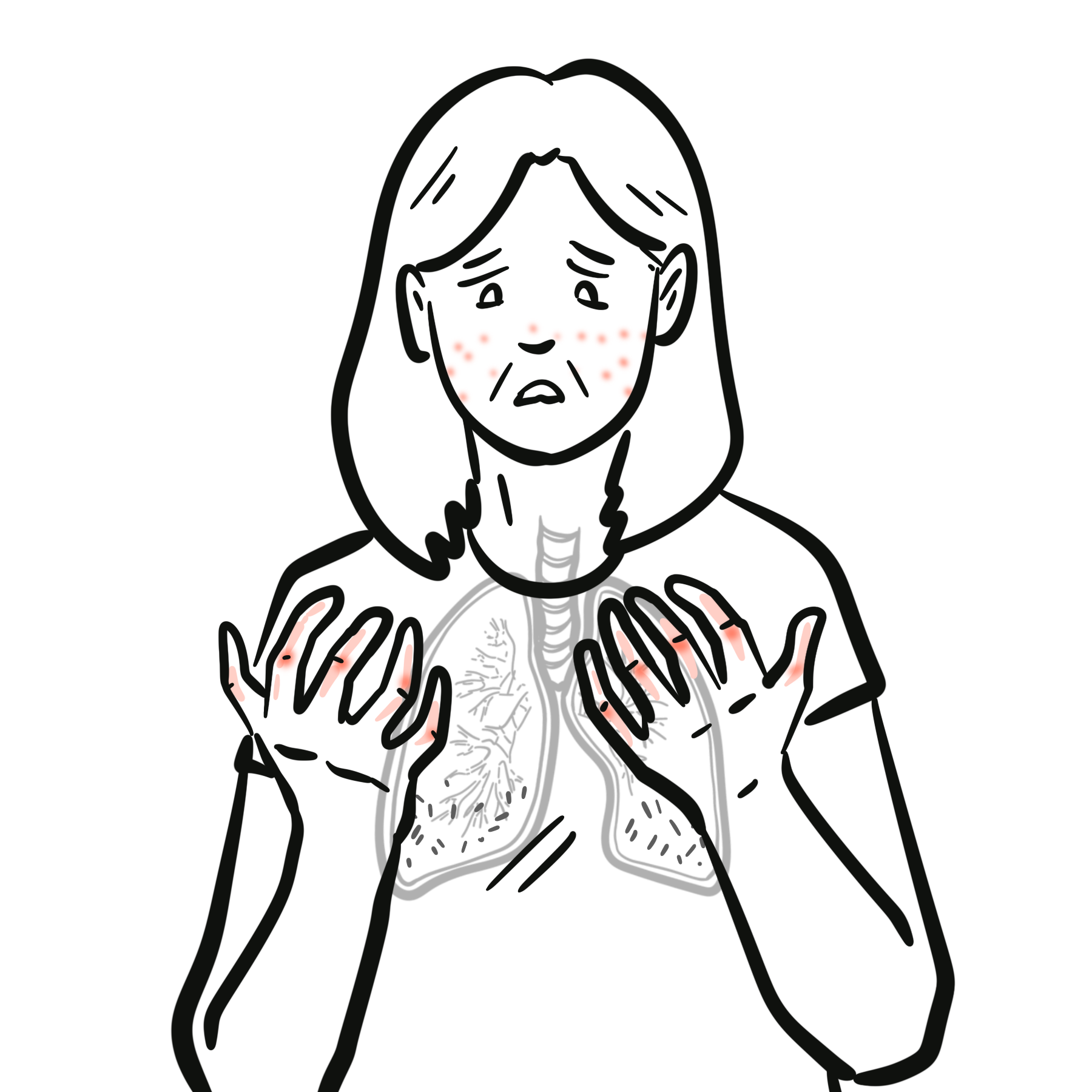
Systemic sclerosis–associated interstitial lung disease (SSc-ILD) is one of the most common and severe internal organ manifestations of systemic sclerosis (SSc), occurring in up to 50–60% of patients on HRCT, with clinically significant disease in ~25–30%. It is a major cause of SSc-related mortality. The most frequent histopathological pattern is nonspecific interstitial pneumonia (NSIP), followed by usual interstitial pneumonia (UIP). Risk factors include diffuse cutaneous SSc, anti-topoisomerase I (Scl-70) antibody positivity, male sex, African-American ethnicity, and early disease onset. The most important complication is progressive pulmonary fibrosis leading to respiratory failure and death.
What to notice
Detailed image description
Definition
Systemic sclerosis (SSc): Chronic autoimmune connective tissue disease characterized by vasculopathy, immune dysregulation, and fibrosis of skin and internal organs.
Interstitial lung disease (ILD): Group of disorders involving inflammation and/or fibrosis of the lung interstitium.
Nonspecific interstitial pneumonia (NSIP): ILD pattern with uniform interstitial inflammation and fibrosis, common in SSc.
Forced Vital Capacity (FVC): Spirometric measure of lung volume, used to monitor ILD progression.
Anatomy & Physiology
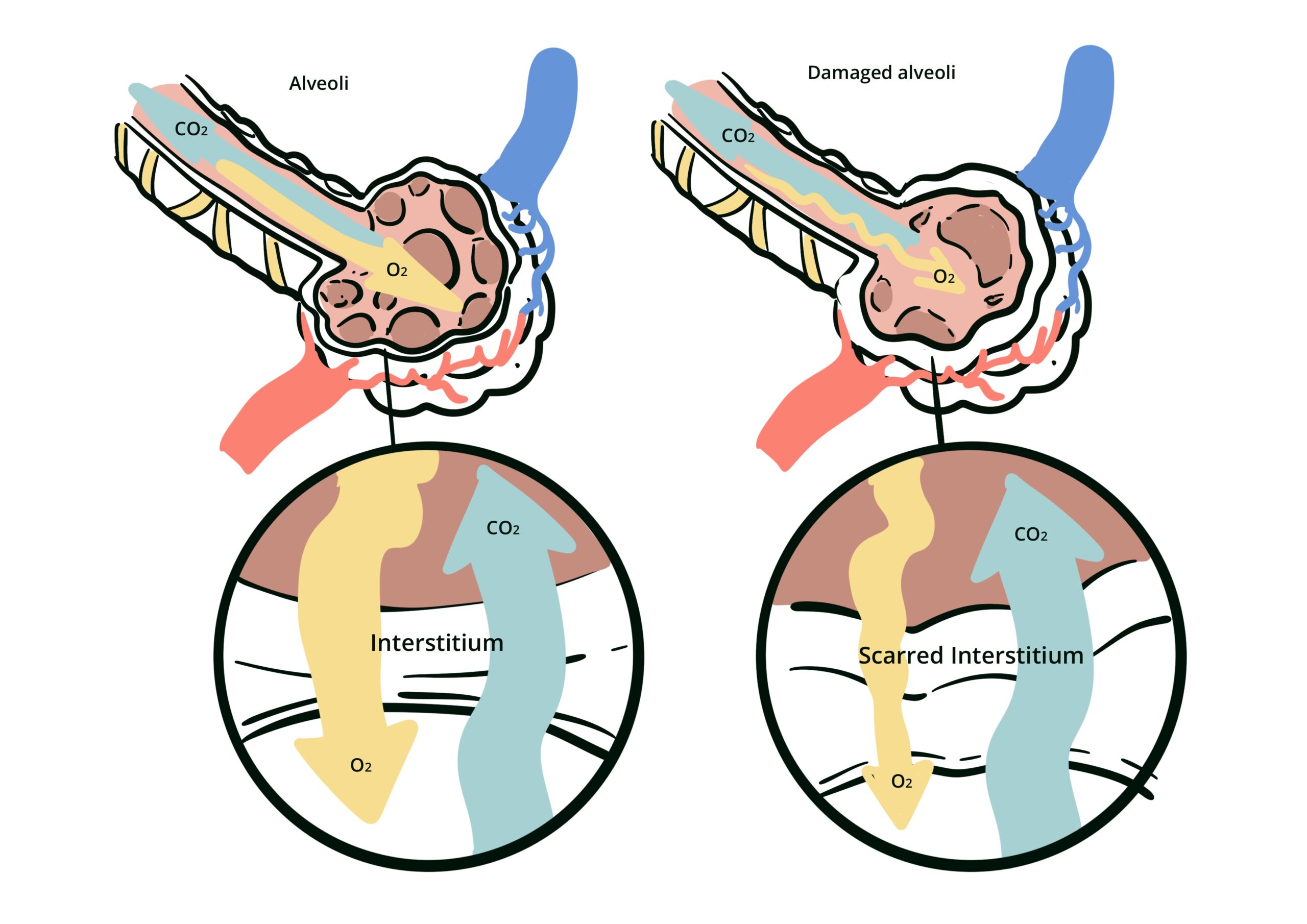
- Alveolar-capillary unit: Site of gas exchange; consists of alveolar epithelium, interstitial space, and capillary endothelium.
- Type I pneumocytes: Thin cells covering most alveolar surface, specialized for gas exchange.
- Type II pneumocytes: Produce surfactant, repair alveolar epithelium after injury.
- Interstitial space: Contains fibroblasts, extracellular matrix; normally thin to allow efficient diffusion.
- Pulmonary microvasculature: Maintains low-resistance blood flow for oxygen uptake.
Aetiology & Risk Factors
Aetiology
- Autoimmune-mediated endothelial injury and fibroblast activation
- Genetic predisposition (e.g., HLA alleles, IRF5, STAT4 variants)
- Environmental exposures (silica, organic solvents)
- Microchimerism (fetal cell persistence in maternal circulation)
Risk Factors
- Diffuse cutaneous SSc subtype
- Anti-Scl-70 antibody positivity
- Male sex
- African-American ethnicity
- Early disease onset (<5 years from SSc diagnosis)
- High baseline CRP or ESR
- Smoking
Anti-centromere antibody is associated with lower ILD risk but higher pulmonary hypertension risk.
Pathophysiology
- Endothelial injury → microvascular dysfunction, increased permeability
- Immune activation → infiltration by T cells, B cells, macrophages
- Cytokine release (TGF-β, IL-6, PDGF) → fibroblast activation
- Fibroblast-to-myofibroblast transition → excessive collagen and ECM deposition
- Interstitial thickening → impaired gas exchange, restrictive lung physiology
- Progressive fibrosis → irreversible architectural distortion, honeycombing (UIP)
Early microvascular injury precedes fibrosis — screening should target early disease phase.
Clinical Manifestations
- Progressive exertional dyspnea
- Nonproductive cough
- Bibasilar “Velcro” crackles
- Fatigue, reduced exercise tolerance
- Digital ulcers, Raynaud’s phenomenon (coexistent SSc features)
- Signs of pulmonary hypertension in advanced disease
Anti-Scl‑70 positivity and diffuse cutaneous SSc are strong predictors of ILD development.
Diagnosis
Diagnostic criteria
- Systemic Sclerosis diagnosis
- HRCT showing ILD pattern
- Exclusion of other ILD causes
- High resolution CT (HRCT)
- NSIP (ground-glass opacities, subpleural sparing)
- UIP (honeycombing, reticulation)
- Pulmonary Function Tests: restrictive pattern, ↓ FVC, ↓ DLCO
- Serology: ANA, anti-Scl-70, anti-centromere, anti-RNA polymerase III
- Echocardiography: screen for pulmonary hypertension
- 6-minute walk test: functional assessment
Differential Diagnoses
| Condition | Differentiating Feature |
| Idiopathic NSIP | No systemic features, negative autoantibodies |
| IPF (UIP) | Older age, no SSc features |
| Hypersensitivity pneumonitis | Exposure history, centrilobular nodules |
| Drug-induced ILD | Temporal relation to drug exposure |
HRCT is more sensitive than CXR; baseline and follow-up imaging are essential.
Classification
- By HRCT/histopathology:
- Nonspecific Interstitial Pneumonia (NSIP) – most common
- Usual Interstitial Pneumonia (UIP)
- Organizing Pneumonia (OP)
- Lymphoid Interstitial Pneumonia (LIP)
- By extent:
- Limited (<20% lung involvement)
- Extensive (>20% lung involvement)
Treatment
- Immunosuppressants:
- Glucocorticoids – not recommended given risk of renal crisis
- Mycophenolate mofetil (first-line)
- Cyclophosphamide OR Rituximab OR Tocilizumab
- Anti-fibrotics:
- Nintedanib (approved for SSc-ILD)
- Supportive:
- Oxygen therapy
- Pulmonary rehabilitation
- Vaccinations (influenza, pneumococcal)
- Monitoring:
- Serial PFTs every 3–6 months
- HRCT as indicated
- Refractory/severe disease
- Autologous stem cell transplant (skin and lung)
- CAR T-cell therapy
Nintedanib slows FVC decline in SSc-ILD regardless of background immunosuppression.
Complications & Prognosis
- Progressive pulmonary fibrosis
- Pulmonary hypertension
- Respiratory failure
- Increased infection risk (immunosuppression-related)
- NSIP: better prognosis than UIP
- Poor prognostic factors:
- Male sex
- African-American ethnicity
- Extensive lung involvement (>20%)
- Rapid FVC decline (>10% in 12 months)
- UIP pattern
Early detection and aggressive management improve survival.
References
- Hoffmann-Vold AM et al. The identification and management of interstitial lung disease in systemic sclerosis: evidence-based European consensus statements. Lancet Rheumatol. 2020;2(2):e71–e83.
- Distler O et al. Nintedanib for systemic sclerosis–associated interstitial lung disease. N Engl J Med. 2019;380:2518–2528.
- Volkmann ER. Natural history of systemic sclerosis–related interstitial lung disease: how to identify a progressive fibrosing phenotype. J Scleroderma Relat Disord. 2020;5(2_suppl):31–40.
- Goh NS et al. Interstitial lung disease in systemic sclerosis: a simple staging system. Am J Respir Crit Care Med. 2008;177:1248–1254.
- Bruni C et al. Predicting ILD in systemic sclerosis: the ILD-RISC model. BMC Pulm Med. 2025;25:3722.
- Khanna D et al. 2013 ACR/EULAR classification criteria for systemic sclerosis. Arthritis Rheum. 2013;65:2737–2747.











Members only discussions coming soon…