

Aspiration Pneumonia
Overview
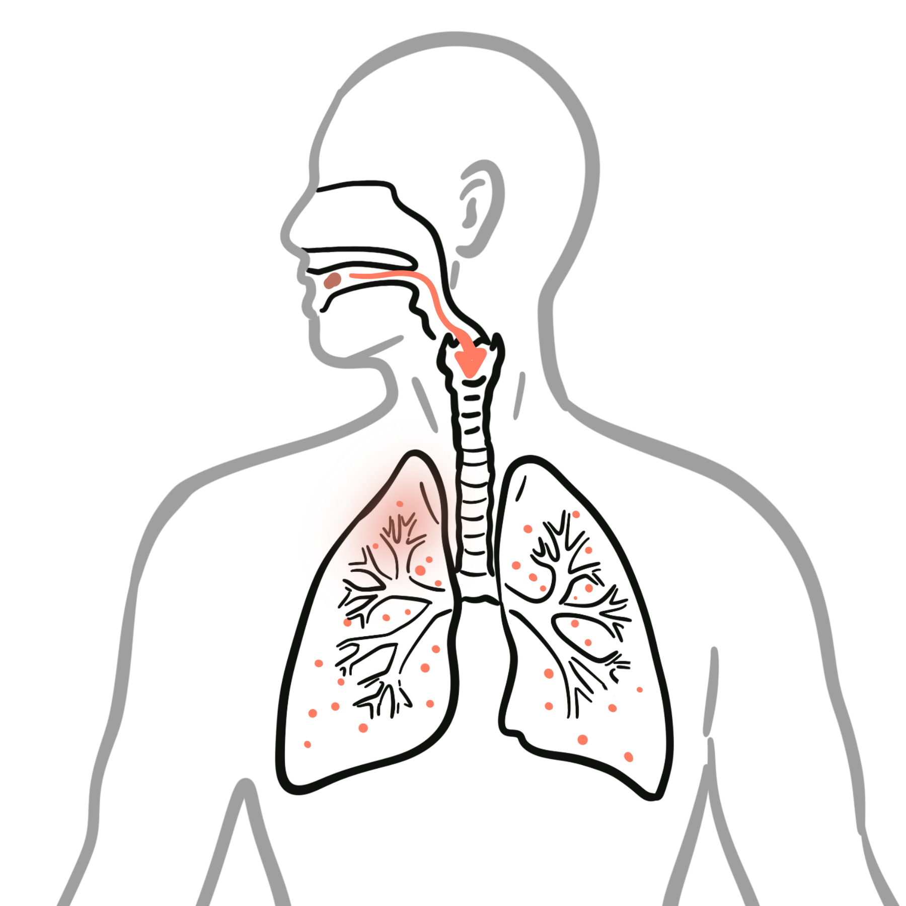
Aspiration pneumonia is an acute pulmonary infection caused by inhalation of bacteria-rich oropharyngeal or upper gastrointestinal material into the lower respiratory tract. It occurs when aspiration is combined with impaired airway protection, ineffective clearance and sufficient pathogenic bacterial burden. It is particularly important in older adults, people with neurological disease, frailty, dysphagia or reduced consciousness.
Aspiration pneumonia must be distinguished from aspiration pneumonitis, a sterile chemical lung injury that usually follows macroaspiration of gastric contents. The two syndromes can overlap and may be difficult to separate at first presentation, but the distinction matters because uncomplicated chemical pneumonitis does not require routine antibiotics.
Severe disease may cause acute hypoxaemic respiratory failure, sepsis, lung abscess or empyema. Immediate priorities are stabilisation, assessment of pneumonia severity and identification of any ongoing aspiration risk.
Definition
- Aspiration pneumonia
- Bacterial infection of the lung following aspiration of colonised oropharyngeal or upper gastrointestinal material.
- Aspiration pneumonitis
- Acute chemical lung injury caused by aspiration of irritant gastric contents, without primary bacterial infection.
- Dysphagia
- Difficulty or impairment in moving food, fluid or saliva safely from the mouth to the stomach.
- Silent aspiration
- Entry of material below the vocal cords without an overt cough or other obvious clinical response.
Anatomy & Physiology
Safe swallowing requires coordinated oral, pharyngeal and oesophageal phases. During the pharyngeal phase, elevation of the larynx, closure of the vocal cords and epiglottic movement protect the airway while the upper oesophageal sphincter relaxes. Sensation within the pharynx and larynx triggers swallowing and cough when material approaches or enters the airway.
Small-volume aspiration occurs even in healthy adults, particularly during sleep. Pneumonia is usually prevented by intact consciousness, glottic closure, an effective cough, mucociliary clearance, immune defence and a relatively low burden of pathogenic oral organisms.
Gravity influences where aspirated material settles. In a supine patient, the posterior upper-lobe segments and superior lower-lobe segments are commonly affected. In an upright patient, the basal lower lobes—often the right—are more exposed. Distribution is not perfectly specific and depends on body position during aspiration.
An aspiration-pattern infiltrate is position-dependent. Ask where and how the patient was positioned during the suspected event before deciding whether the radiograph supports aspiration.
Aetiology & Risk Factors
Aetiology
Aspiration pneumonia usually follows aspiration of colonised oral or pharyngeal secretions. The microbiology resembles other community- or healthcare-associated pneumonias more closely than the historically assumed purely anaerobic infection. The likely pathogens depend on where the pneumonia was acquired, recent healthcare exposure, prior antimicrobial therapy and individual risk factors for resistant organisms.
Less commonly, aspiration of infected gastric contents may occur in the presence of gastric bacterial colonisation, intestinal obstruction or enteral feeding. Repeated aspiration may produce recurrent pneumonia or contribute to chronic suppurative airway disease.
Risk Factors
Risk is highest when impaired swallowing or airway protection is combined with poor oral health and reduced physiological reserve.
- Oropharyngeal dysphagia: stroke, Parkinson disease, dementia, motor neurone disease, multiple sclerosis, cerebral palsy or other neuromuscular disease
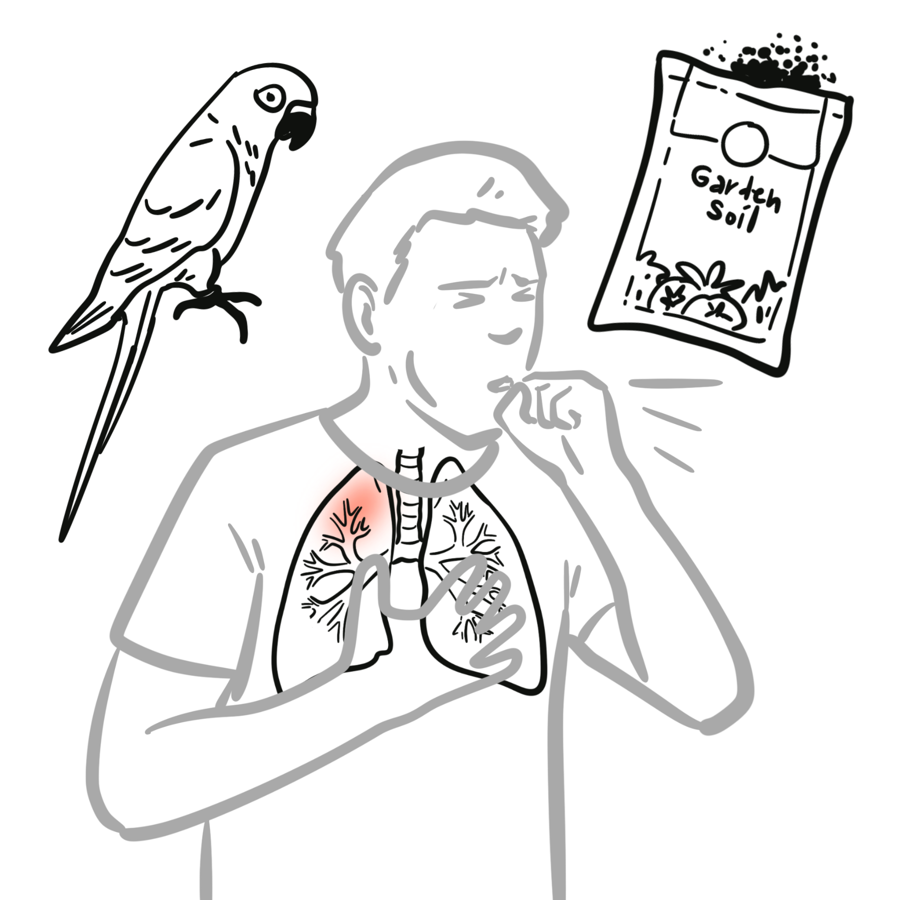
- Reduced consciousness: alcohol, sedatives, opioids, seizures, anaesthesia, head injury, metabolic encephalopathy or critical illness
- Impaired cough or airway clearance: frailty, immobility, respiratory muscle weakness, tracheostomy or chronic lung disease
- Structural or treatment-related swallowing impairment: head and neck cancer, radiotherapy, oesophageal disease, recent intubation or upper gastrointestinal surgery
- Poor oral health: dental caries, periodontal disease, xerostomia and dependence on others for oral care
- Feeding-related factors: dependence for feeding, rushed feeding, inappropriate positioning or poorly fitted dentures
- Gastro-oesophageal factors: reflux, vomiting, gastroparesis or bowel obstruction
- General vulnerability: advanced age, malnutrition, dehydration, immunosuppression and multimorbidity
Enteral feeding does not abolish aspiration risk. Patients may still aspirate oral secretions or refluxed gastric contents, and feeding tubes should not be regarded as automatic protection against pneumonia.1,2
Many aspiration events are unwitnessed. The absence of choking or vomiting does not exclude aspiration pneumonia, particularly in frail adults or people with neurological disease and silent aspiration.
Pathophysiology
Aspiration delivers bacteria and inflammatory material beyond the larynx. Whether pneumonia develops depends on the volume and composition of the aspirate, bacterial burden, site of deposition and the patient’s cough, mucociliary and immune defences. Impaired clearance allows organisms to proliferate, producing alveolar inflammation, consolidation and ventilation–perfusion mismatch.
The inflammatory response causes fever, cough, sputum production, hypoxaemia and radiographic infiltrates. Severe infection may extend locally to cause necrosis, lung abscess or pleural infection, or systemically to cause sepsis and multiorgan dysfunction.
Aspiration pneumonitis follows a different initial mechanism: acidic or otherwise irritant gastric contents cause direct epithelial injury and an abrupt inflammatory response. Symptoms may begin within hours of a witnessed macroaspiration and can improve substantially with supportive care over the next 24–48 hours. Secondary bacterial infection can subsequently occur, particularly when clinical deterioration or failure to improve persists.
A witnessed aspiration event followed by abrupt hypoxaemia and bilateral infiltrates may be chemical pneumonitis rather than bacterial pneumonia. Stabilise first, then reassess the clinical trajectory; antibiotics are not automatically required for uncomplicated pneumonitis.
Clinical Manifestations
Presentation ranges from a typical acute pneumonia to a subtle decline in a frail older adult. A witnessed aspiration event is helpful but is not required.
Stroke, seizures and alcohol impair swallowing and airway protection, while vomiting increases aspiration risk
Features suggesting ongoing aspiration
- Recurrent pneumonia, particularly with dependent-lobe involvement
- Coughing, choking or breathlessness during eating or drinking
- Prolonged mealtimes, food pocketing or multiple swallows
- Wet or gurgling voice after swallowing
- Unexplained weight loss, dehydration or recurrent low-grade fever
- New swallowing difficulty after stroke, intubation or neurological deterioration
Aspiration pneumonitis more often presents with abrupt respiratory distress, hypoxaemia, bronchospasm and diffuse infiltrates shortly after a clear macroaspiration. Aspiration pneumonia more often evolves over hours to days with infective features, although substantial overlap limits certainty at presentation.
Diagnosis
Aspiration pneumonia is a clinical diagnosis supported by evidence of pneumonia together with aspiration risk or a compatible aspiration event. No single test confirms the diagnosis.
Initial assessment
- Measure oxygen saturation, respiratory rate, heart rate, blood pressure, temperature and conscious state.
- Assess sepsis and pneumonia severity using clinical judgement and a locally accepted tool such as CURB-65 where appropriate; frailty, comorbidity and baseline function may add important information not captured by a score.
- Obtain an arterial or venous blood gas when there is significant hypoxaemia, respiratory distress, altered consciousness or concern about ventilatory failure or lactate.
Focused investigations
- Chest radiograph: first-line imaging
- Lower lobes, especially right
- Full blood count, urea, electrolytes and renal function: assess inflammatory response, dehydration, renal impairment and factors affecting antimicrobial choice or dosing.
- C-reactive protein
- Blood cultures: reserve for severe pneumonia, sepsis
- Sputum microscopy and culture: useful in severe disease, treatment failure, recurrent infection, bronchiectasis, recent healthcare exposure or risk of resistant pathogens; a poor-quality sample is difficult to interpret.
In an upright patients, the right lung is more commonly affected because the right main bronchus is wider and more vertical.
In a supine patient, posterior upper lobes and superior lower lobes are typical sites.

Image: melvil via Wikimedia Commons, licensed under CC BY-SA 4.0.
When to escalate imaging or procedures
- CT chest: use when the chest radiograph is inconclusive despite strong clinical concern, when an obstructing lesion or foreign body is possible, or when complications such as abscess, necrosis or empyema are suspected.
- Pleural ultrasound and diagnostic aspiration: perform when a clinically significant pleural effusion is present and pleural infection is possible.
- Bronchoscopy: not routine; consider for suspected foreign-body aspiration, airway obstruction, persistent lobar collapse or failure to respond despite appropriate therapy.
Swallowing assessment
Once the patient is stable, screen for dysphagia and refer to speech pathology when aspiration risk is suspected. A bedside assessment identifies risk and informs immediate feeding precautions, but it cannot reliably exclude silent aspiration. Instrumental assessment with videofluoroscopic swallow study or fibreoptic endoscopic evaluation of swallowing is appropriate when the mechanism remains uncertain or detailed information would alter rehabilitation and feeding decisions.1
| Feature | Aspiration pneumonia | Aspiration pneumonitis |
|---|---|---|
| Typical event | Often unwitnessed; recurrent microaspiration | Often witnessed macroaspiration of gastric contents |
| Onset | Evolves over hours to days | Abrupt, often within hours |
| Dominant features | Fever, purulent sputum, pneumonia syndrome | Hypoxaemia, bronchospasm, respiratory distress |
| Imaging | Dependent-segment infiltrate; focal or multifocal | Often bilateral or diffuse dependent infiltrates |
| Initial treatment | Antibiotics plus supportive care | Supportive care; antibiotics only if bacterial infection is suspected or emerges |
Neither fever, leukocytosis nor an infiltrate proves bacterial infection immediately after macroaspiration: chemical injury can cause all three. Time course and response over the next 24–48 hours often provide the most useful discrimination.
Treatment
Initial management
- Oxygen if hypoxic
- Airway protection if necessary
- Suction/clear secretions
- IV fluids if septic/dehydrated
- Assess swallowing and aspiration risk
Antibiotics
For established aspiration pneumonia, choose antibiotics according to local guidelines and severity.
Common approaches include:
- Amoxicillin–clavulanate for community-acquired aspiration pneumonia
- Severe/hospital-acquired cases may require broader coverage depending on local resistance patterns.
- Anaerobic coverage is particularly considered when there is lung abscess or empyema, rather than being automatically required for every aspiration event.
“Aspiration” does not automatically mean “anaerobes.” Treat according to the pneumonia setting and resistance risk; add specific anaerobic coverage when abscess, empyema or necrotising infection makes it clinically relevant.
Manage swallowing, nutrition and oral health
- Arrange speech pathology assessment and implement individualised swallowing strategies.
Aspiration pneumonitis
Provide supportive respiratory care after chemical aspiration. Antibiotics are not routinely indicated when the clinical picture is consistent with uncomplicated pneumonitis and the patient improves promptly. Reassess closely and initiate or continue antibiotics when bacterial pneumonia cannot be excluded, clinical deterioration occurs or improvement fails to emerge.
Recurrent aspiration pneumonia is often a systems problem: unsafe swallowing, poor positioning, inadequate feeding assistance, oral bacterial burden and sedating medicines may act together. Prevention is therefore multidisciplinary.
Complications & Prognosis
Complications
- Lung abscess
- Empyema
- Necrotising pneumonia
- Respiratory failure
- ARDS
- Bronchiectasis following recurrent aspiration
- Septic shock
Aspiration pneumonia + persistent fever + cavitary lesion → think lung abscess.
Prognosis
Outcome is influenced by pneumonia severity, age, frailty, neurological disease, level of consciousness, nutritional status, comorbidity and whether the aspiration risk can be modified. Aspiration pneumonia generally carries a worse prognosis than non-aspiration community-acquired pneumonia because affected patients often have impaired swallowing, poor functional reserve and multiple comorbidities.1,2
Clinical improvement should be evident within the first few days of appropriate treatment. Failure to improve should prompt review of the diagnosis, antimicrobial adequacy, resistant organisms, continued aspiration, airway obstruction and complications such as abscess or empyema.
Recurrent episodes warrant reassessment of swallowing, oral care, feeding support, medication burden and goals of care. In advanced neurodegenerative disease or severe frailty, management should include realistic discussion of prognosis, burdens of intervention and the patient’s preferences.1
References
- Simpson AJ, Allen JL, Chatwin M, Crawford H, Elverson J, Ewan V, et al. BTS clinical statement on aspiration pneumonia. Thorax. 2023;78(Suppl 1):s3–s21. doi:10.1136/thorax-2022-219699
- Mandell LA, Niederman MS. Aspiration pneumonia. N Engl J Med. 2019;380(7):651–663. doi:10.1056/NEJMra1714562
- Metlay JP, Waterer GW, Long AC, Anzueto A, Brozek J, Crothers K, et al. Diagnosis and treatment of adults with community-acquired pneumonia: an official clinical practice guideline of the American Thoracic Society and Infectious Diseases Society of America. Am J Respir Crit Care Med. 2019;200(7):e45–e67. doi:10.1164/rccm.201908-1581ST
- Bai AD, Srivastava S, Digby GC, Girard V, Razak F, Verma AA. Anaerobic antibiotic coverage in aspiration pneumonia and the associated benefits and harms: a retrospective cohort study. Chest. 2024;166(1):39–48. doi:10.1016/j.chest.2024.02.025







Members only discussions coming soon…