

Orthostatic Hypotension
Overview
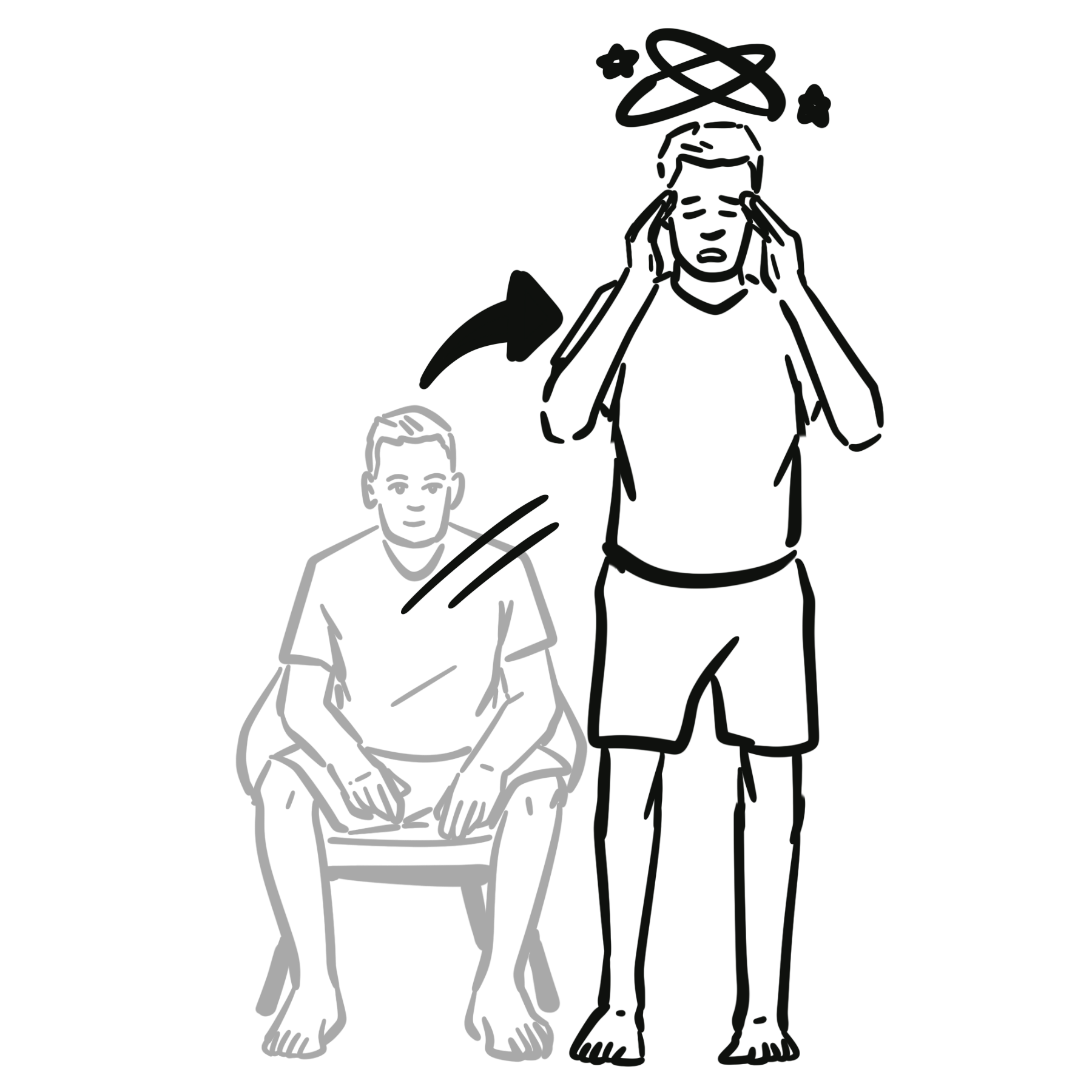
Orthostatic Hypotension (OH) is defined as a sustained drop in blood pressure occurring within 3 minutes of standing or head-up tilt.
- Systolic BP: Decrease of >= 20 mmHg AND/OR
- Diastolic BP: Decrease of >= 10 mmHg
(In patients with supine hypertension >= 150/90 mmHg, a drop of >= 30 mmHg SBP is required).
Normal Physiological Response to Orthostasis (standing up)
- Standing up from a recline or sitting position results in pooling of 500-1000mL of blood in the lower extremities and splanchnic circulation.
- The physiological response
- ↓venous return to the heart → ↓ventricular filling → ↓cardiac output → baroreceptor reflex
- Baroreceptor reflex
- → stimulation of the vasomotor center → peripheral vasoconstriction → ↑BP → ↑venous return + ↑cardiac output
- → stimulation of the cardiovascular center → ↑HR → ↑cardiac output +↑venous return
- Overall clinically there is a small reduction in systolic BP (<10mmHg), increase in diastolic BP and an increase in HR
Always measure blood pressure after at least 5 minutes of quiet rest in the supine position before performing an active stand test to establish a true baseline.
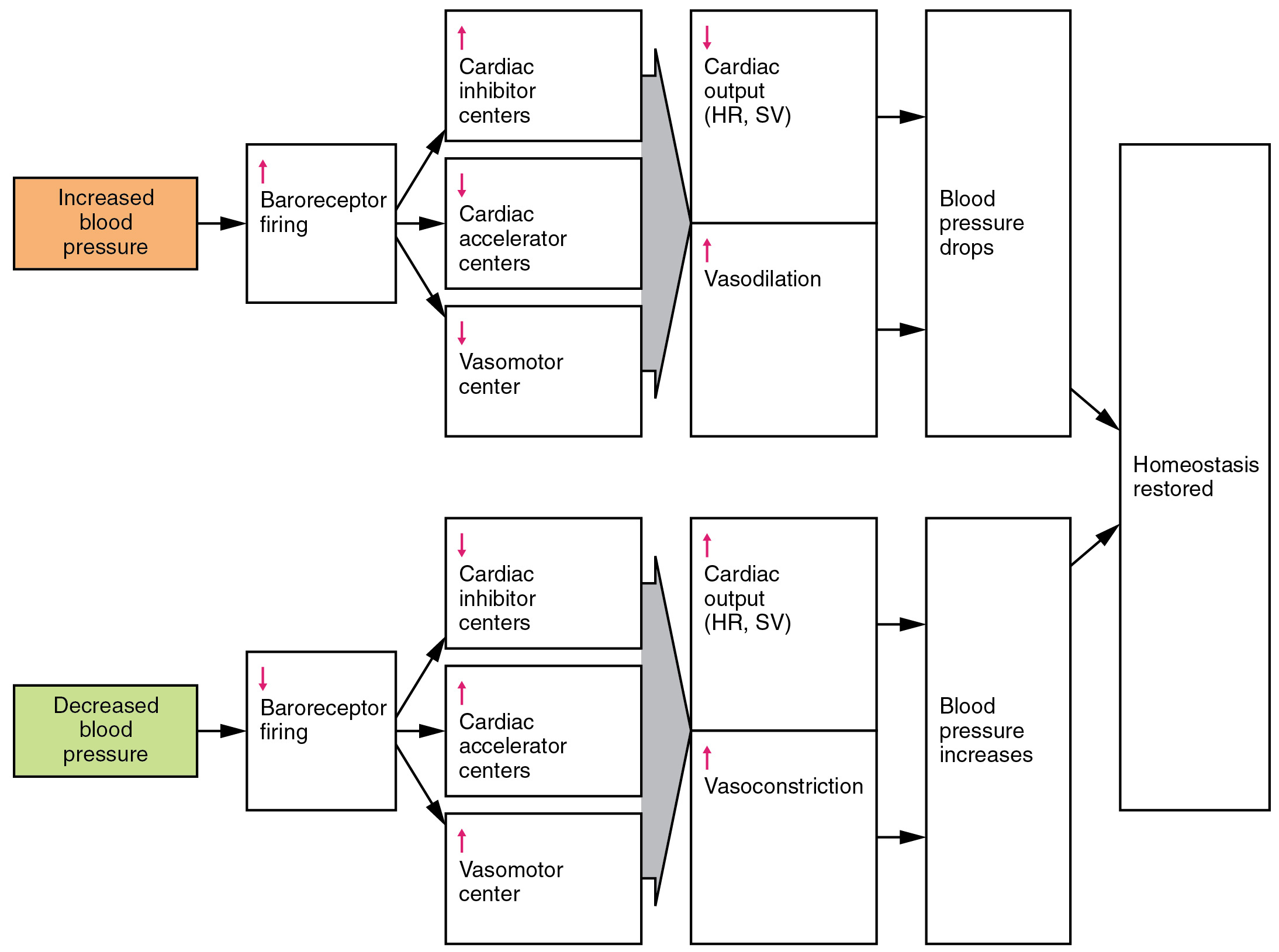
For orthostasis, follow the lower pathway: falling blood pressure reduces baroreceptor firing, increasing cardiac accelerator and vasomotor activity while reducing cardiac inhibition. Heart rate, contractility and vascular tone rise to support blood pressure. The upper pathway shows the opposite response to a rise in pressure.
What to notice
Detailed image description
Two negative-feedback pathways are shown. Increased pressure increases baroreceptor firing, promotes cardiac inhibition and reduces accelerator and vasomotor activity, lowering cardiac output and dilating vessels. Decreased pressure reduces baroreceptor firing, reduces cardiac inhibition and increases accelerator and vasomotor activity, raising cardiac output and constricting vessels. Both pathways restore pressure towards baseline.
Triage & Approach
Step 1: Diagnostic Confirmation (Active Stand Test / Bedside Orthostatics)
- Measurement Protocol: Measure HR and BP after 5 minutes resting supine, then at 1 minute and 3 minutes post-standing.
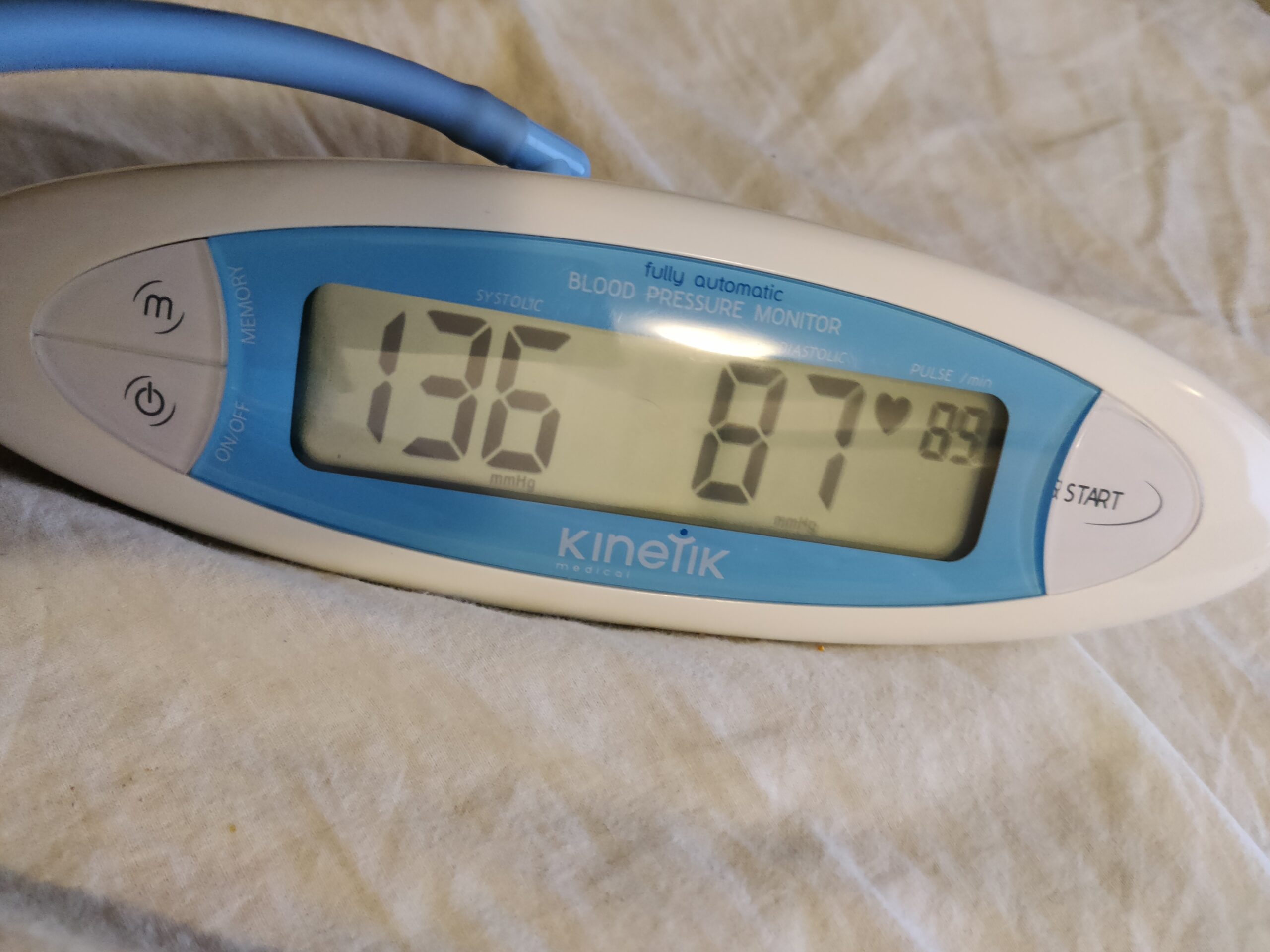
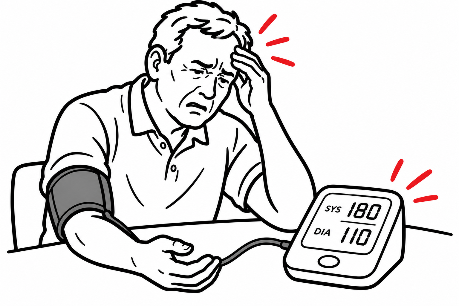
Record systolic pressure, diastolic pressure and heart rate at each stage of the active stand test: after at least 5 minutes supine, then at 1 and 3 minutes standing. This photograph illustrates the three readouts; a single displayed measurement does not diagnose orthostatic hypotension. Support the cuffed arm at heart level and supervise standing for safety.
What to notice
Detailed image description
An automated blood-pressure monitor displays systolic and diastolic pressure in mmHg and pulse in beats per minute. Orthostatic assessment compares sequential readings taken in the specified positions, rather than interpreting one isolated reading.
Step 2: Emergency Triage & Red Flags
- Syncope with Trauma or Head Injury: Rule out intracranial hemorrhage/cervical spine injury.
- Acute Volume Loss / Hemorrhage: Tachycardia + severe orthostasis -> GI bleed, ruptured ectopic pregnancy, ruptured AAA.
- Acute Cardiac Signs: Chest pain, dyspnea, irregular pulse -> Arrhythmia, ACS, aortic stenosis, PE.
- Neurological Red Flags: Rapidly progressive parkinsonism, ataxia, early autonomic failure, vertical gaze palsy -> Central neurodegenerative etiologies (e.g., Multiple System Atrophy).
Step 3: Branching Logic (Heart Rate Response Analysis)
- Neurogenic Orthostatic Hypotension (nOH): Blunted HR increase (< 15 bpm upon standing despite significant BP drop). Indicates baroreflex arc failure or sympathetic denervation.
- Non-Neurogenic Orthostatic Hypotension: Compensatory reflex tachycardia (> 15 bpm HR increase upon standing). Indicates hypovolemia, vasodilation, or drug-induced etiology.
- POTS Differential: HR increase >= 30 bpm (or >= 40 bpm if age 12–19) within 10 minutes of standing WITHOUT a drop in SBP >= 20 mmHg.2
A blunted heart rate response (< 15 bpm increase despite a > 20 mmHg drop in SBP) strongly points toward a neurogenic etiology (autonomic failure), whereas prominent compensatory tachycardia indicates volume depletion or medication-induced vasodilation.
Differential Diagnosis
Non-Neurogenic Causes (Intact Baroreflex, Compensatory Tachycardia):
- Volume Depletion / Hypovolemia:
- Distinguishing features: Dehydration, vomiting/diarrhea, overt GI bleeding, excessive diuresis, elevated BUN/creatinine ratio (> 20:1), dry mucous membranes.
- Medication-Induced (Most common overall cause in elderly):
- Distinguishing features: Antihypertensives (alpha-blockers e.g., tamsulosin/doxazosin, vasodilators, diuretics), psychotropics (TCAs, antipsychotics), SGLT2 inhibitors, nitrates.
- Cardiovascular Deconditioning / Prolonged Bedrest:
- Distinguishing features: Recent prolonged hospitalization, frailty, muscle wasting; intact autonomic reflexes.
- Adrenal Insufficiency (Addison’s Disease):
- Distinguishing features: Hyperpigmentation, hyponatremia, hyperkalemia, weight loss, fatigue, hypoglycemia.
Neurogenic Causes (Impaired Baroreflex, Blunted HR Response):
- Central Synucleinopathies (Primary Autonomic Failure):
- Parkinson’s Disease (PD): Resting tremor, rigidity, bradykinesia + orthostasis.
- Multiple System Atrophy (MSA): Orthostatic hypotension + prominent ataxia (MSA-C) or early severe autonomic failure with erectile dysfunction and urinary retention (MSA-P/Shy-Drager).
- Dementia with Lewy Bodies (DLB): Visual hallucinations, fluctuating cognition, parkinsonism + early orthostasis.
- Pure Autonomic Failure (PAF): Isolated autonomic failure without central neurological signs.
- Peripheral Neuropathies (Secondary Autonomic Failure):
- Diabetic Autonomic Neuropathy: Longstanding diabetes, stocking-glove sensory loss, gastroparesis, resting tachycardia.
- AL Amyloidosis: Nephrotic syndrome, macroglossia, periorbital purpura (“raccoon eyes”), sensorimotor neuropathy + severe orthostasis.
- Autoimmune Autonomic Ganglionopathy (AAG): Acute/subacute severe pan-autonomic failure (fixed pupils, dry mouth, severe orthostasis; anti-gAChR antibodies).
- Other Neuropathies: Sjögren’s syndrome, HIV, Vitamin B12 deficiency, Guillain-Barré / Miller Fisher variant.
Tamsulosin and other alpha-1 blockers used for BPH are notorious causes of drug-induced orthostatic hypotension in older men. Always review the medication list before initiating an extensive neurological workup.
Investigations
First-Line / Bedside & Basic Diagnostic Tests:
- Active Stand Test (Bedside Orthostatics): Standard 3-minute test quantifying SBP, DBP, and HR changes.
- 12-Lead ECG: Assess for arrhythmia, ischemic changes, conduction delay, or prolonged QTc.
- Full Blood Count (FBC): Screen for anemia (occult bleeding/hemolysis).
- Urea, Electrolytes, Creatinine: Screen for dehydration (elevated BUN/Cr), renal dysfunction, or Addisonian electrolyte shifts (low Na+, high K+).
- Fasting Glucose & HbA1c: Rule out diabetic neuropathy / metabolic causes.
- Morning Cortisol (8 AM): Screen for adrenal insufficiency.
Targeted / Specialized Autonomic Investigations:
- Head-Up Tilt Table Testing (HUTT): Indicated if active stand test is inconclusive, or to differentiate nOH from POTS or vasovagal syncope.
- Autonomic Function Tests:
- Ewing Battery: Heart rate variability with deep breathing (30:15 ratio), Valsalva maneuver phase IV overshoot, cold pressor test.
- Serum Free Light Chains, SPEP/UPEP: Screen for AL Amyloidosis / Plasma cell dyscrasia.
- Serum Anti-Neuronal Ganglionic Acetylcholine Receptor (gAChR) Antibodies: Confirm Autoimmune Autonomic Ganglionopathy.
- Supine and Standing Plasma Norepinephrine Levels:
- Normal: Norepinephrine doubles upon standing.
- nOH (Central/PAF): Low supine norepinephrine (< 100 pg/mL) with minimal rise upon standing (< 100% increase).
In normal individuals, standing causes plasma norepinephrine levels to double. A failure of norepinephrine to rise upon standing confirms neurogenic autonomic failure.
Critical Management
Immediate Stabilization (Symptomatic Acute / Severe Drops):
- Positioning: Place patient supine with legs elevated (Trendelenburg position) to optimize venous return and cerebral perfusion.
- Acute Fluid Bolus: If hypovolemic or acutely symptomatic without heart failure -> Rapid infusion of 500 mL normal saline IV (or rapid ingestion of 500 mL cold water orally, which triggers a sympathetically mediated pressor response lasting 30–60 min).
- Drug Re-evaluation: Hold or depressurize non-essential antihypertensives, diuretics, and alpha-blockers immediately.
First-Line Non-Pharmacological Measures (Core Long-Term Strategy):
- Physical Counterpressure Maneuvers (PCM): Leg crossing, muscle squeezing, squatting, or toe-raising at onset of symptoms.
- Dietary Sodium & Fluid Intake: 2–3 L fluid/day + 6–10 g salt/day (unless contraindicated by severe heart failure).
- Abdominal Binder & Waist-High Compression Stockings: Abdominal compression (15–20 mmHg) is more effective than lower leg compression alone due to splanchnic venous pooling.
The splanchnic circulation holds up to 20–30% of total blood volume. An abdominal binder is often far more effective at preventing orthostatic drops than thigh-high compression stockings alone.1,2
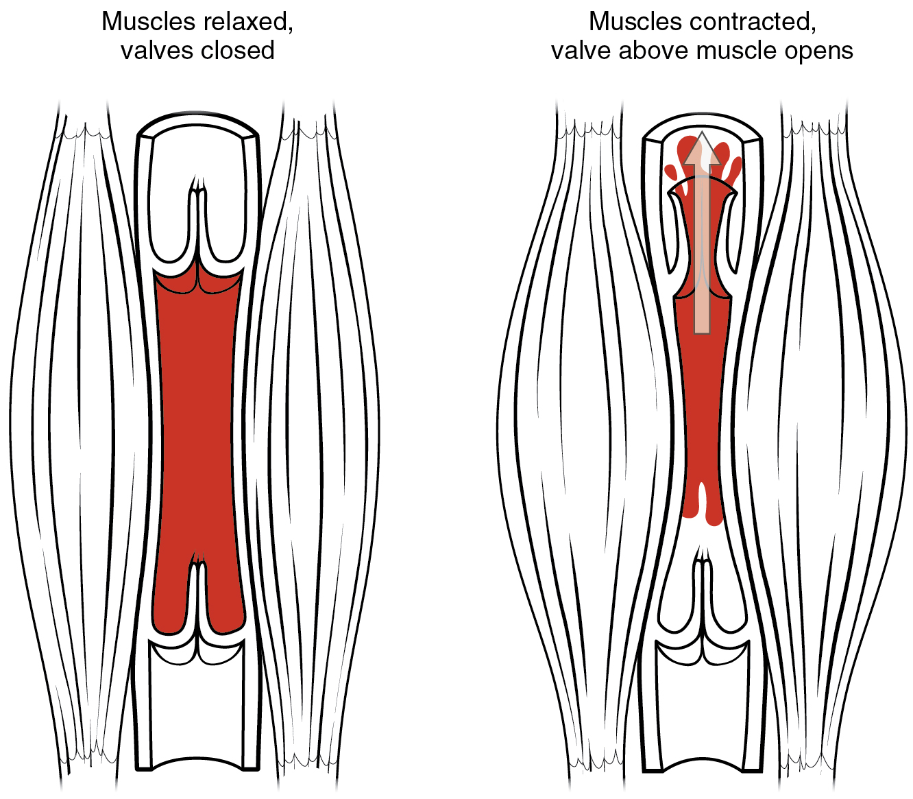
Compare the relaxed and contracted muscle panels. Muscle contraction compresses the vein and drives blood towards the heart, while one-way valves limit backflow. This mechanism helps explain why leg-muscle activation can reduce venous pooling and support the counterpressure manoeuvres described above.
What to notice
Detailed image description
The left panel shows relaxed skeletal muscles around a vein. The right panel shows contracting muscles compressing the vein: the valve above the contraction opens and blood moves upwards towards the heart, while the lower valve prevents reverse flow.
References
- Gibbons CH, Schmidt P, Biaggioni I, Frazier-Mills C, Freeman R, Isaacson S, et al. The recommendations of a consensus panel for the screening, diagnosis, and treatment of neurogenic orthostatic hypotension and associated supine hypertension. J Neurol. 2017;264(8):1567-1582. doi:10.1007/s00415-016-8375-x
- Kim MJ, Farrell J. Orthostatic hypotension: a practical approach. Am Fam Physician. 2022;105(1):39-49. Available from: https://www.aafp.org/afp/2022/0100/p39
Image sources and licences
- Baroreceptor reflex: compensation for a fall in blood pressure. OpenStax College, Anatomy & Physiology, via Wikimedia Commons; reproduced without alteration. CC BY 3.0.
- Bedside assessment: record blood pressure and pulse together. Kent Madsen, via Wikimedia Commons; reproduced without alteration. CC BY-SA 4.0.
- Skeletal muscle pump: supporting venous return. OpenStax College, Anatomy & Physiology, via Wikimedia Commons; reproduced without alteration. CC BY 3.0.


Members only discussions coming soon…