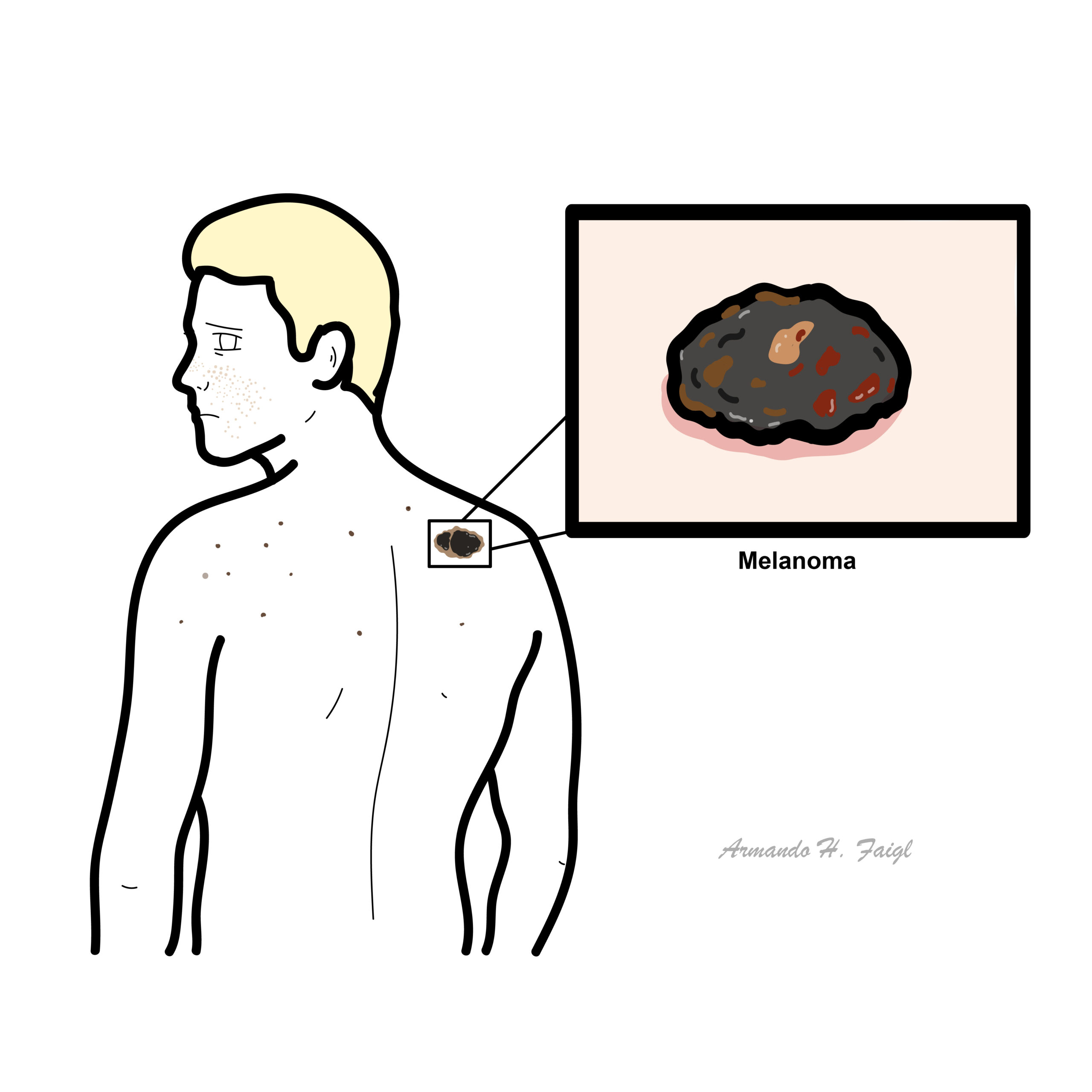
My Account
auto-complete
more

Field of Study

Field of Study

Post Type

Post Type

Melanoma

Notes »
disease
» Dermatology
Melanoma

Overview

Overview Melanoma accounts for 4 percent of all dermatological cancer. Melanoma has the highest mortality rate of all dermatological cancers, only 14 percent of patients with metastatic melanoma survive for five years. Melanoma is one of the most common cancers in young adults.

Video: Melanoma Overview

Anatomy and Physiology

The skin consist of three layers, from superficial to deep:

  • Epidermis
  • Dermis
    • Papillary layer
    • Reticular layer
  • Hypodermis (some don't classify it as skin)

The epidermal layer are made up mainly of keratinocytes, which are squamous epithelial cells high in keratin (protein). Other cells residing in the epidermis include:

  • Melanocytes - produce melanin
  • Langerhan cells - immune cell, basically a dendritic cell of the skin
  • Merkel Cells - tactile cells for sensation

Melanocytes are mature melanin-forming cells found in many parts of the body (eye, ear, hair, heart, bone), but especially in the skin. Through a process called melanogenesis, these cells produce melanin, which is a pigment found in the skin, eye and hair. Vitamin D and UV light stimulate melanocytes to produce melanin.

Watch Dermatology Introduction

Risk Factors

Riskfactors

Signs and Symptoms

  • Atypical naevi
  • Ulceration and bleeding
  • Constitutional symptoms

Examination - ABCDE

  • A - asymmetry
  • B - borders (irregular and/or distinct)
  • C - colour (varied)
  • D - Diameter (increasing or >6mm)
  • E - Elevation and Evolution (most important as in changing rapidly/growing)
Remember Blanching. Press down on the lesion for ~3 seconds. Release if pale and refills there is a vascularity involved.

Diagnosis

  • Examination ABCDE
  • If Melanoma suspected perform a skin biopsy
  • Breslow's thickness can be used to stage Melanoma and establish prognosis

Differential Diagnosis

  • Seborrhoeic keratosis
  • Pigmented basal cell carcinoma
  • Actinic keratosis
  • Benign melanocytic naevi
  • Spitz naevus
  • Haemangioma
Think Squamous cell carcinoma is not a good differential because it is not pigmented.

Investigations

  • Dermatoscope
  • Skin Biopsy
  • Chest X-ray for metastasis
Remember 1.4cm depth is dangerous.

Pathophysiology

  • Melanoma arises from melanocytes
  • Genetics and environmental factors play a role in the pathogenesis
  • A proportion of melanoma arise from pre existing naevi

Pathophysiology

Mutated BRAF and NRAS genes are most common in superficial spreading and nodular melanoma.

Pathology

Different types of Melanoma

  • Superficial spreading (most common)
  • Nodular (most dangerous)
  • Lentigo maligna
  • Acral lentiginous
Pathological Differences between Benign and Malignant tumours
MalignantBenign
InvasiveNon-Invasive
Poorly differentiatedWell Differentiated
High mitotic rateNormal mitotic rate
Non-capsulatedCapsulated
Usually large growthUsually small growth
Not well circumscribedWell circumscribed

Staging

Clark's Staging - Important histologically
Clark's LevelHistological characteristic
IConfined to the epidermis "in situ"
IIInvasion of the papillary dermis
IIIFilling of the papillary dermis
IVInvasion of the reticular dermis
VInvasion of the deep, subcutaneous tissue
Breslow's Staging - Important Prognostically
Breslow's ThicknessDepth of tumour invasion
I≤0.75mm
II0.75-1.5mm
III1.51-2.25mm
IV2.26-3.0mm
V>3.0mm

Management

  • Surgical excision
  • Topical Imiquimod
  • Surgical excision +/- sentinel lymph node biopsy

Complications and Prognosis

Complication

Metastasis

Treatment adverse effects

  • Chemotherapy
  • Radiotherapy
  • Immunotherapy (causes flu-like symptoms)

Prognosis The most important prognostic indicators are Breslow's depth of the tumour and lymph node status.

  • Stage 0 (in-situ melanoma): >98%
  • Stage I (Breslow's depth <1 mm and no nodal or metastatic disease): 90% to 95%
  • Stage II (localised disease, intermediate to thick depth): 45% to 78% (78% for non-ulcerated melanoma of depth 1 to 4 mm; 45% for an ulcerated melanoma >4 mm depth)
  • Stage III (nodal metastases): 69% (non-ulcerated melanoma of any depth with a single positive node) to 26% (ulcerated melanoma of any depth with 4 or more positive nodes)
  • Stage IV (metastatic): overall prognosis is bleak; 3% to 10% depending on the extent and sites of metastasis.

References

UpToDate
RACGP
Best Practice
Coming soon
Note taking just got easier!
Take a note easily on any page.
Refer to your notes on the page they were taken
Access all note in your profile. Easily navigate to the relevant page.
Download your note at any time.
More new features coming soon.
Get updates on new releases.
hello world!
envelopemagnifiercross
0
Would love your thoughts, please comment.x
()
x