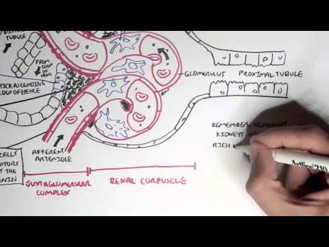
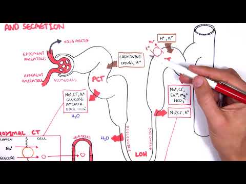
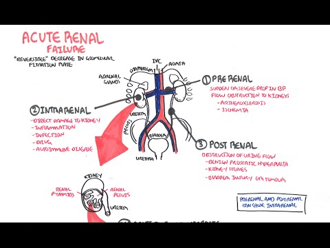
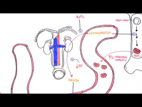
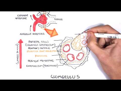
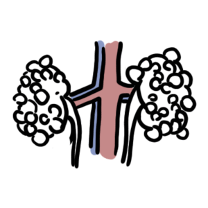
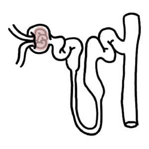
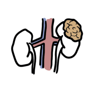
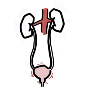
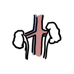
0:00 Armano Hasudun, Biology and Medicine videos. Please make sure to subscribe. 0:06 Join the 0:06 four main group for the latest videos. Please visit Facebook, Armano Hasudun 0:10 and 0:10 here you can like. Please ask questions, answer some questions and post some 0:14 interesting things such as artworks. It'd be greatly appreciated and you can 0:18 also change the quality settings to the highest one for better graphics. And in 0:22 this video we're going to talk about nephrology, about the kidneys, the 0:25 nephrons, etc. And so we have this person with an open abdominal cavity and 0:31 this 0:31 is where the kidneys are situated as well as other functional organs. Now if we 0:38 zoom into the abdominal here, the open abdominal, we can see two kidneys. We 0:43 have 0:43 each two kidneys. The left one makes a lot slightly higher than the right and 0:47 above the kidneys we have adrenal glands and these adrenal glands secrete 0:51 hormones. Now the kidneys, a normal function is to produce urine which then 0:56 travels through the ureter into the urethra where it can then be expelled by 1:01 our body either through the penis or the vagina. Now also we have the arteries, 1:07 the 1:08 big aorta which supplies our kidneys and others organs around it and then we 1:13 have the inferior vena cava which brings the blood back to the heart. Now 1:18 let's zoom into one of these kidneys and look at its anatomy briefly. So we're 1:25 looking at the left kidney here with a cross-sectional view. So we know that 1:29 the 1:29 kidney produces urine but why does it produce urine? Well it does this because 1:33 the kidneys regulate the pH, the volume and composition of the blood and so it 1:39 also eliminates nitrogenous waste via urine. And it also has another function 1:45 in that it secretes a particular hormone, erythropoietin which I will not 1:49 discuss in this video. Now the main structures of the kidneys, we have the 1:53 renal cortex, the outer part section of the kidney and then we have the renal 1:59 medulla, the inner part of the kidney and then we have the renal pyramids of 2:04 this 2:05 of the medulla and pyramids are only situated in the medulla usually. And then 2:10 we have the renal pelvis which connects to the ureter. So again the kidneys has 2:16 a 2:16 superficial cortex and a deeper medulla consisting mainly of medulla 2:22 pyramids. And now the kidneys have a very rich blood supply because this is 2:29 how the kidneys regulate the blood pH, volume and composition. Now the the 2:35 artery which brings blood into the kidneys is called the renal artery. The most 2:40 important thing about the kidney is that they contain what's called nephrons 2:43 which are situated in the renal cortex and in the renal medulla. And these 2:48 nephrons are the functional unit of kidneys and we have many many many 2:53 nephrons about one and a one and a half million per kidney. And the and the 2:59 nephrons are the ones that actually filtrate, reabsorb and secrete substances 3:04 to make urine to be expelled by the body. And so they are the ones that help 3:11 regulate blood pH, composition and volume. So nephrons are the big deal. Now 3:17 here you can see this is the head of the nephron and it continues down to form 3:23 a 3:23 loop and it forms a loop within the medulla, the renal medulla and as you can 3:28 see here's a renal cortex. And we have blood supply coming into the head of the 3:37 nephrons. Because blood begins getting filtered in the head of the nephron 3:40 which then all the substances which have been filtered and travels through the 3:45 nephron through here following this route and then out through this long 3:51 vertical tube known as a collecting duck. Now let's just concentrate on the 3:58 blood 3:58 supply again to the kidneys. Remember that the aorta is the big artery which 4:05 then will form the renal artery which supplies the kidneys. The blood will then 4:12 travel to essentially the afferent arterials at the very end. And the 4:18 afferent arterials is what goes into this head of the nephron. Known as the 4:23 renal 4:24 corpuscle which consists of the glomerular capsule, the outer part of the head 4:27 and 4:28 most importantly the glomerulus that is a really difficult word to say. And the 4:34 glomerulus is formed from the afferent arterials and this is where filtration 4:39 occurs. And then after this the glomerulus will form the efferent arterials 4:44 which 4:44 leaves the nephron essentially. This is the blue thing. Now the function of the 4:51 nephron as I mentioned is for filtration, tubular reabsorption and tubular 4:57 secretion which occurs all along the nephron except filtration which occurs 5:02 at the glomerulus. So let's just say when the afferent arterials brings blood 5:09 to 5:09 the renal corpuscle, the glomerulus will then do filtration and then all the 5:16 substances and water will then travel down through the nephron. The first part 5:22 it travels down through is called the proximal convoluted tubule which then 5:27 travels to the loop of Henley, this big loop. And the loop of Henley consists 5:31 of 5:31 two parts, the descending loop and the ascending loop of Henley. The ascending 5:36 loop of Henley will then form the distal convoluted tubules where the blood 5:42 will 5:42 still be traveling. The distal convoluted tubule actually has to loop back to 5:49 where the glomerulus was, the renal corpuscle. And we'll see the reason for 5:54 this later on. And essentially then the distal coveted tubule will then connect 5:59 to this vertical long tube known as a collecting duct which will then 6:03 essentially connect to the ureter which will then expel the urine out of the 6:07 kidneys, out of the body. Now back to this diagram where the 6:12 efferent arterials brings blood back out of the head of the nephron. It then 6:17 can 6:17 form the vasorecta or it can form the renal vein essentially and then will form 6:24 the inferior vena cava. Now this vasorecta only occurs, only is present in 6:32 certain types of nephrons. And we'll get back to this soup. But another 6:38 important 6:38 thing to remember is that each part of the nephron contains different types of 6:42 cells. And so let's just draw another diagram of these nephrons. Now here I 6:50 will 6:50 draw two types of nephrons because there are two main types of nephrons. One 6:55 which 6:56 consists of the vasorecta, the vein, the special vein. Now this long-looking ne 7:03 phron 7:03 which dips down into the medulla is known as the juxta medullary nephron. 7:09 And it has a long loop of Henley and it consists and it contains the vasorecta 7:14 vessels which I mentioned previously. And this other one, the short-looking one 7:19 , 7:19 which just dips to the medulla but is more prominent in the cortex is known as 7:24 a cortical nephron. And it has a short loop of Henley and doesn't go deep into 7:28 the medulla. Remember the blood travels towards the renal corpuscle which will 7:36 then, where we'll then get filtrated in the glomerulus and all these substances 7:41 and water will then travel through the nephron and reabsorption and secretion 7:47 will occur which will then form urine. And this urine will then travel down 7:51 through the to the collecting duct into the ureter which will then be 7:55 expelled by the body. Okay now I mentioned this earlier but kidneys have very 8:01 rich 8:02 blood supply and this is important in order to regulate the blood composition, 8:06 pH, volume, and to eliminate waste as well as for reabsorption and secretion 8:13 all along the nephron. And so basically the kidneys has a rich blood supply and 8:19 this is because once the effort arterial leaves the renal corpuscle from the 8:28 glomerulus, it will then begin wrapping around and basically traveling all 8:35 through the nephron, all along the nephron. And this is in order to enable the 8:41 nephron to perform reabsorption and secretion all along the nephron. And 8:46 once the the nephron has been reabsorbing and secreting all its 8:50 substances into this capillary, the capillary will then form the vein which 8:55 will then form this bigger vein and essentially will form the renal vein and 8:58 then will, yeah, continue on. Now the juxtaposed a medullary nephron is 9:06 different to the cortical nephron in that it has a vasorecta and the vasorecta 9:10 is also the capillary light structure which is formed after the 9:17 efferent arterials, after the efferent arterial leaves the renal corpuscle. And 9:22 what's important about vasorecta is that it's sort of like a straight vessel 9:29 and this enables some serious secretion and reabsorption of water down this big 9:37 loop of handling. And so actually the vasorecta and juxtaposed a medullary 9:42 nephron are important in establishing the medullary osmotic gradient and so 9:47 helps in essentially in water balance and to either make the urine concentrated 9:54 or 9:54 not as concentrated, you can say. Finally we can look at the different types of 9:59 cells all along the nephron. Let's begin with the collecting duct here and they 10:04 consist of two types of cells mainly the cuboidal cells, the principal cells 10:09 and 10:09 the intercalating cells. And then let's look at the loop of handling. The loop 10:14 of 10:14 handling has these thin segmented cells which are which which sort of epit 10:19 helial 10:19 like cells. So it's very good for reabsorption and secretion. And then let's 10:24 go to the distal convoluted tubules which consists of just your regular 10:28 cuboidal cells. And this can also perform reabsorption and secretion. And then 10:34 we 10:34 have the proximal convoluted tubule cells which are also cuboidal cells. But 10:40 what's unique about them is that they have micro vili on the surface. And 10:43 micro vili are like finger-like projections which help in reabsorption. 10:48 And that's what they're important for. And then we have the renal corpusll 10:53 which 10:53 consists of the two main things, the glomerular capsule and the glomerulus 10:59 cell. Now the glomerular capsule contains epithelial like cells, very thin 11:04 prior 11:04 prior to cells. And the glomerulus contains these special cells known as 11:09 podocytes. And they are attached onto basically the basement membrane of the 11:15 glomerulus. Okay now let's zoom into the renal corpusll here to put some of 11:21 these 11:22 cells to context the podocytes, the parietal cells for example. So here we 11:26 have the blood vessel coming into the renal corpusll. The afferent 11:32 arterioles brings in blood and other substances into to form the glomerulus. 11:38 And then surrounding it we have the glomerular capsule made up of parietal 11:43 cells here. And then these substances in the glomerulus will then get 11:48 filtered by these special cells called podocytes which are on the basement 11:53 membranes. And this area here is known as a renal corpusll where all this 11:58 occurs. 11:58 And then the glomerulus and the renal corpusll will then filtrate all these 12:04 substances and bring it towards the proximal convoluted tubules which are 12:09 made up of these cuboidal like cells with micro vili on the top. And once 12:15 filtration has occurred within the glomerulus and this sort of capsule like 12:20 a structure the bollman's capsule but we'll talk about that later on. The 12:25 glomerulus will then form the efferent arterioles which will then bring blood 12:30 out. And so the glomerulus all which forms a bollman's capsule which I'll talk 12:38 about in the next video is for filtration. In the next video we're going to 12:41 talk 12:42 about the physiology of the nephrons and all other interesting stuff that 12:49 occurs there. Hope you enjoyed it. Please like comment and please provide 12:53 feedback if possible. Thank you very much.