

Alcoholic Cardiomyopathy
Overview
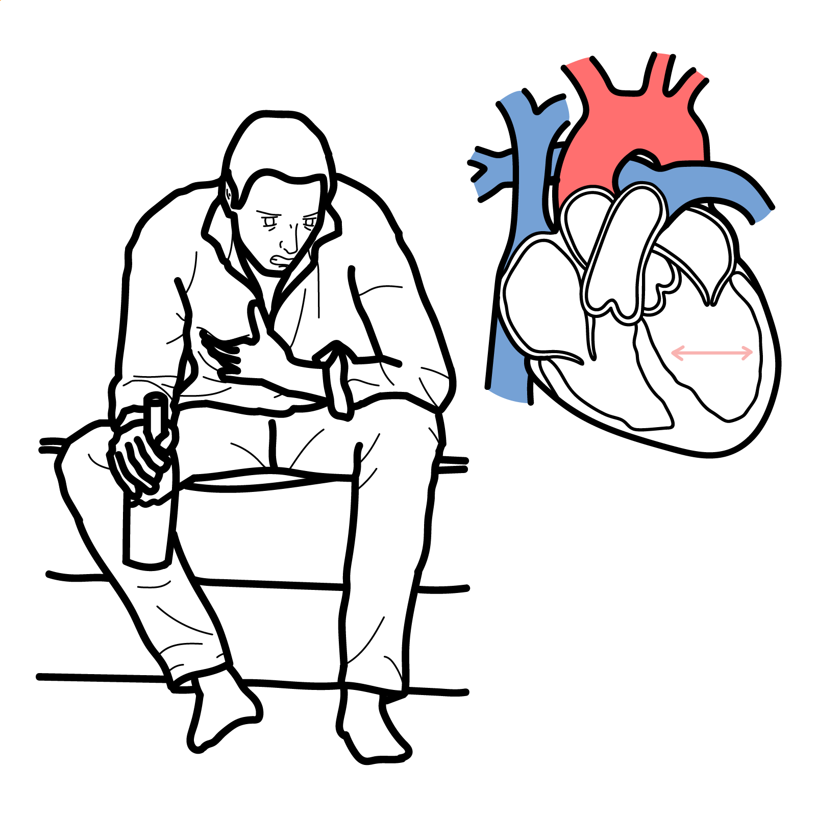
Alcoholic cardiomyopathy, increasingly termed alcohol-induced cardiomyopathy, is an acquired form of dilated cardiomyopathy caused by prolonged heavy alcohol exposure in the absence of another sufficient cause. It typically produces left ventricular dilatation and systolic dysfunction, leading to heart failure, arrhythmias, thromboembolism and sudden cardiac death.1,2
The commonly used exposure threshold is more than 80 g of alcohol daily for at least five years. However, this is a conventional research definition rather than an absolute biological threshold: susceptibility varies with sex, genetics, drinking pattern, nutrition and coexisting cardiovascular disease.1
Early recognition is important because abstinence can produce substantial improvement or recovery of left ventricular function. Continued heavy alcohol consumption is associated with progressive heart failure and substantially worse outcomes.1
Definition
- Alcohol-induced cardiomyopathy
- An acquired dilated cardiomyopathy attributed to prolonged heavy alcohol exposure after other important causes have been excluded.
- Dilated cardiomyopathy
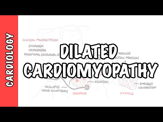
- Left or biventricular dilatation with systolic dysfunction that is not explained solely by abnormal loading conditions or coronary artery disease.
- Standard drink
- A unit containing a defined quantity of pure alcohol; its alcohol content varies between countries, so intake should be recorded in grams rather than drinks whenever possible.
- Reverse remodelling
- Improvement in ventricular size and systolic function following removal of myocardial injury and effective treatment.
Anatomy & Physiology
The ventricles generate pressure and stroke volume through coordinated myocardial contraction. Cardiac output depends on heart rate and stroke volume, which is influenced by preload, afterload and myocardial contractility.
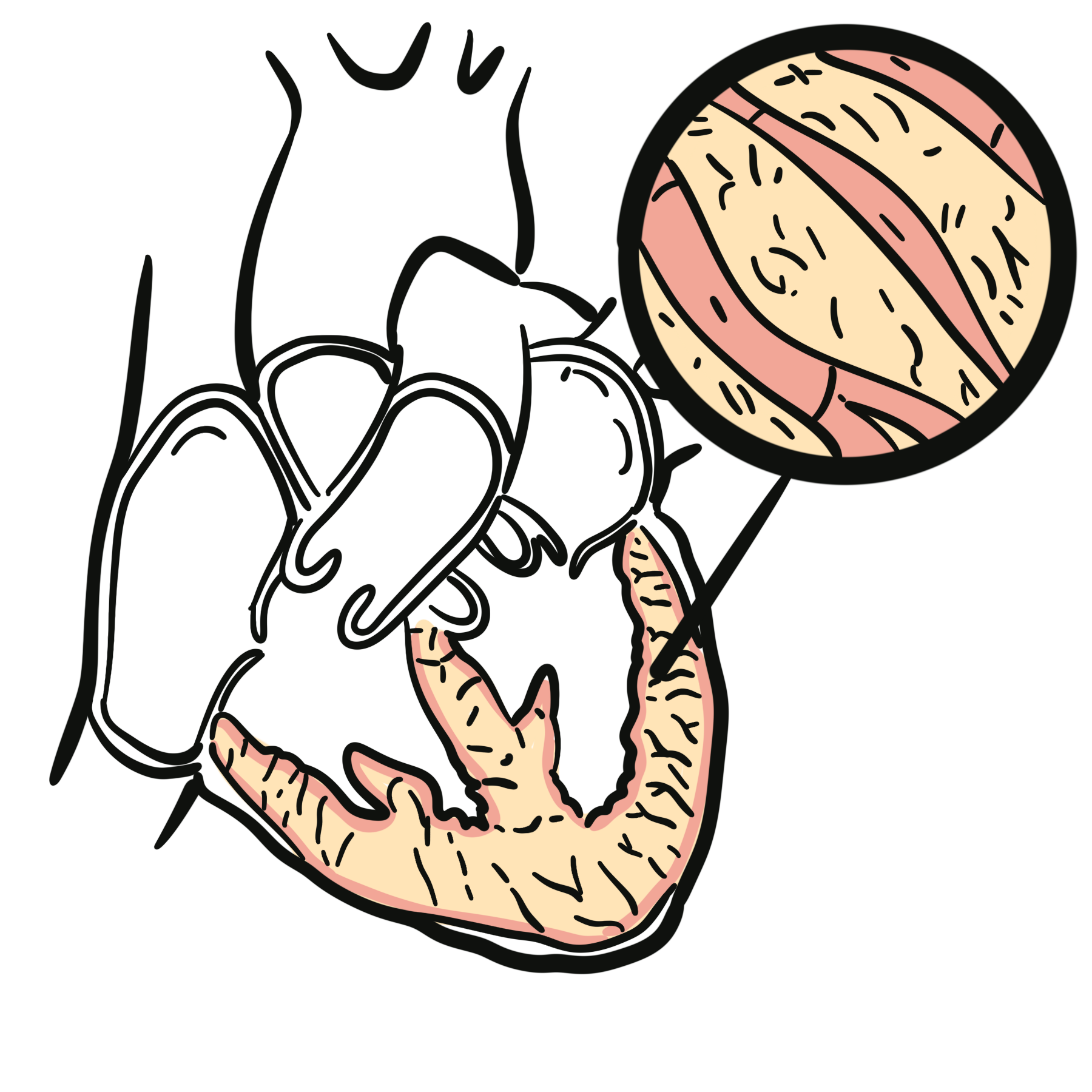
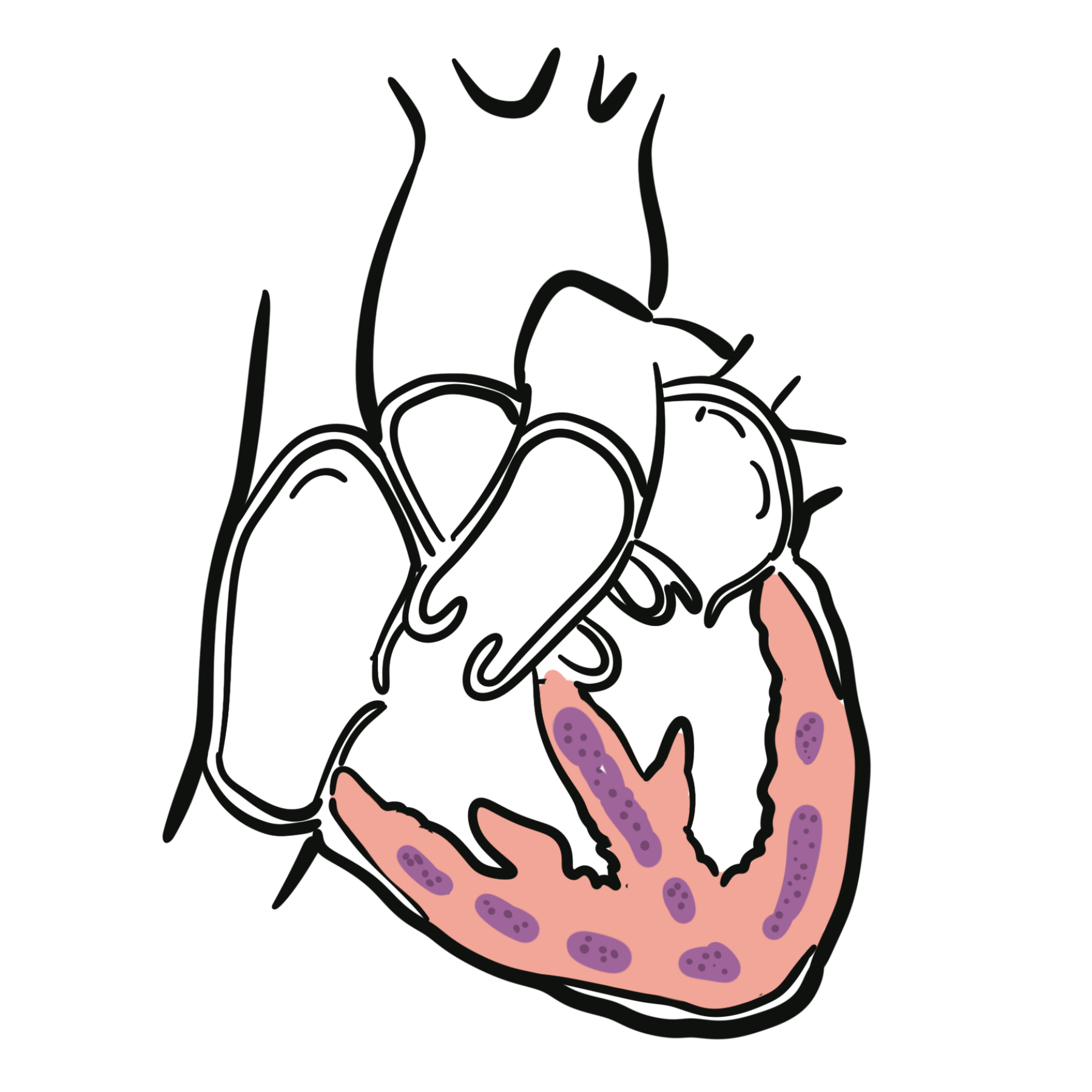
The left ventricle normally ejects blood into the systemic circulation while maintaining an efficient chamber shape and wall thickness. Ventricular dilatation initially helps preserve stroke volume through the Frank–Starling mechanism. Persistent dilatation, however, increases wall stress, worsens mitral regurgitation and reduces mechanical efficiency.
Aetiology & Risk Factors
Aetiology
Alcohol-induced cardiomyopathy results from the cumulative myocardial toxicity of ethanol and its metabolites. It is generally diagnosed when a dilated cardiomyopathy phenotype occurs in the setting of prolonged heavy alcohol consumption and no more convincing cause is identified.1
Alcohol may be the primary cause or one contributor within a multifactorial cardiomyopathy. Pathogenic variants associated with dilated cardiomyopathy—particularly truncating variants in the TTN gene—have been identified in some affected patients, supporting a genetic–environmental “double-hit” model.1,2
Risk Factors
- prolonged heavy daily alcohol consumption
- recurrent binge drinking
- longer duration and greater cumulative alcohol exposure
- female sex, because myocardial toxicity may occur at a lower cumulative exposure
- genetic susceptibility to dilated cardiomyopathy
- hypertension
- nutritional deficiency, particularly thiamine deficiency
- liver disease
- tobacco, cocaine or other cardiotoxic substance exposure
- previous chemotherapy or another myocardial toxin
- coexisting atrial fibrillation or persistent tachycardia
- poor adherence to heart-failure treatment.
The absence of the conventional threshold of more than 80 g/day for five years does not exclude alcohol-related myocardial injury. Conversely, exceeding that threshold does not prove causation.1
Quantify alcohol exposure in grams per day and establish the duration and drinking pattern. The number of “drinks” alone is unreliable because standard-drink definitions and serving sizes vary.
Pathophysiology
Chronic ethanol exposure and acetaldehyde accumulation cause oxidative stress, mitochondrial dysfunction, impaired calcium handling and disruption of excitation–contraction coupling. Alcohol may also impair actin–myosin interaction, alter protein synthesis and activate neurohormonal pathways.1
Progressive myocyte injury and apoptosis reduce myocardial contractility. Compensatory sympathetic and renin–angiotensin–aldosterone activation initially supports cardiac output but promotes sodium retention, vasoconstriction, fibrosis and adverse ventricular remodelling.
The ventricle becomes dilated and increasingly spherical. Functional mitral regurgitation, rising filling pressures and reduced forward output then produce the clinical syndrome of heart failure. Electrical remodelling, chamber enlargement and alcohol’s direct electrophysiological effects increase the risk of atrial and ventricular arrhythmias.
Alcohol exposure does not act in isolation. A genetically susceptible myocardium or coexisting hypertension may lower the exposure required to produce ventricular dysfunction, explaining why patients with similar alcohol histories can have very different cardiac outcomes.1,2
Clinical Manifestations
There are no symptoms or examination findings that reliably distinguish alcohol-induced cardiomyopathy from other forms of dilated cardiomyopathy.1
Patients may report:
- exertional dyspnoea
- reduced exercise tolerance
- fatigue or weakness
- orthopnoea
- paroxysmal nocturnal dyspnoea
- peripheral swelling
- abdominal distension or early satiety
- palpitations
- dizziness, presyncope or syncope
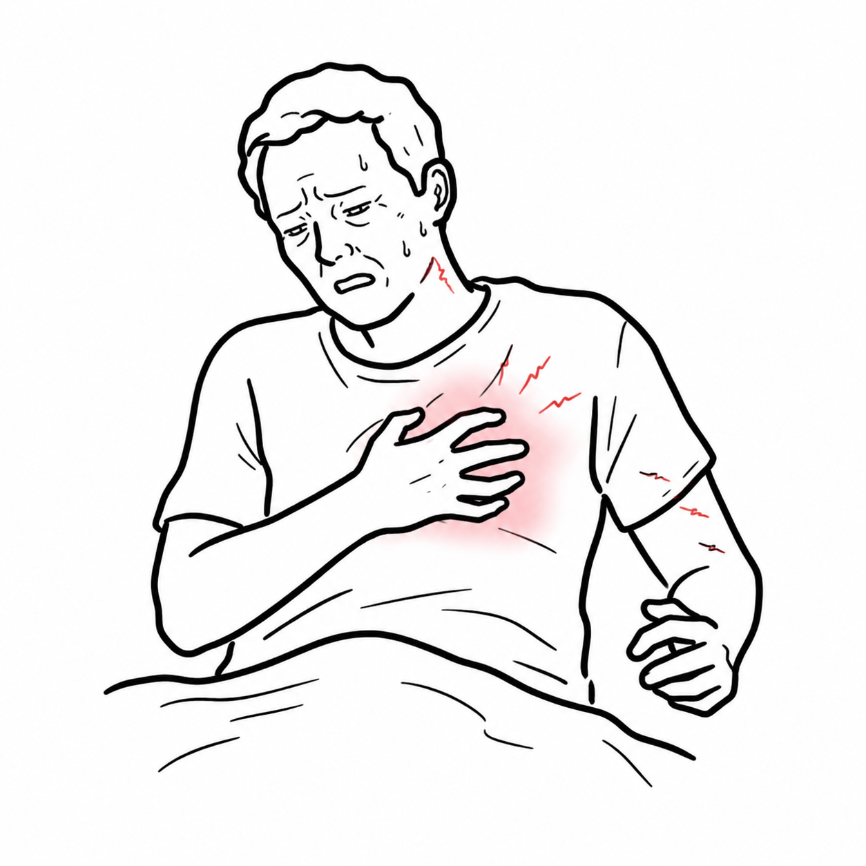
- chest discomfort.
Early disease may be asymptomatic and detected through an abnormal electrocardiogram or cardiac imaging.
Clinical Examination
- tachycardia or an irregular pulse
- hypotension or a narrow pulse pressure in advanced disease
- elevated jugular venous pressure
- displaced, diffuse apex beat
- a third heart sound
- a pansystolic murmur from functional mitral regurgitation
- basal pulmonary crackles
- peripheral oedema
- hepatomegaly or ascites
- cool peripheries and impaired perfusion in advanced heart failure.
Associated alcohol-related findings may include malnutrition, tremor, peripheral neuropathy, hepatic disease, cognitive impairment or features of alcohol withdrawal.
Urgent assessment is required for acute pulmonary oedema, cardiogenic shock, sustained ventricular arrhythmia, syncope, severe electrolyte disturbance or suspected acute coronary syndrome.
Diagnosis & Investigations
Alcohol-induced cardiomyopathy is a diagnosis of attribution and exclusion. A commonly used definition comprises:
- prolonged heavy alcohol consumption, conventionally more than 80 g/day for at least five years
- left ventricular dilatation exceeding two standard deviations above normal
- left ventricular ejection fraction below 50%
- exclusion of other important causes of dilated cardiomyopathy, including coronary, hypertensive and valvular heart disease.1
These criteria are useful for consistency but should not be interpreted as a perfect diagnostic test. No biomarker or imaging pattern is specific for alcohol-induced cardiomyopathy.
Initial assessment generally includes:
- 12-lead electrocardiogram
- transthoracic echocardiogram
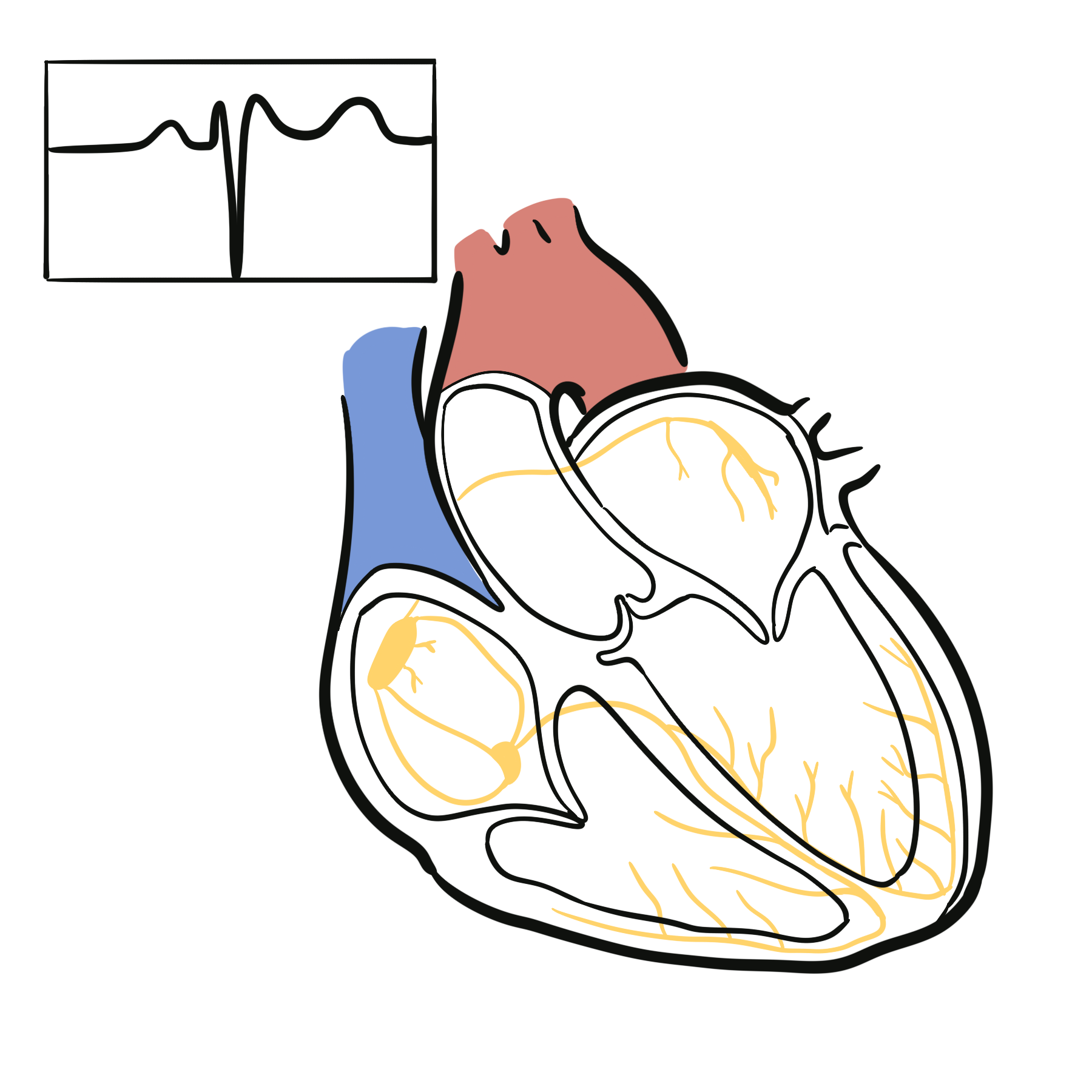
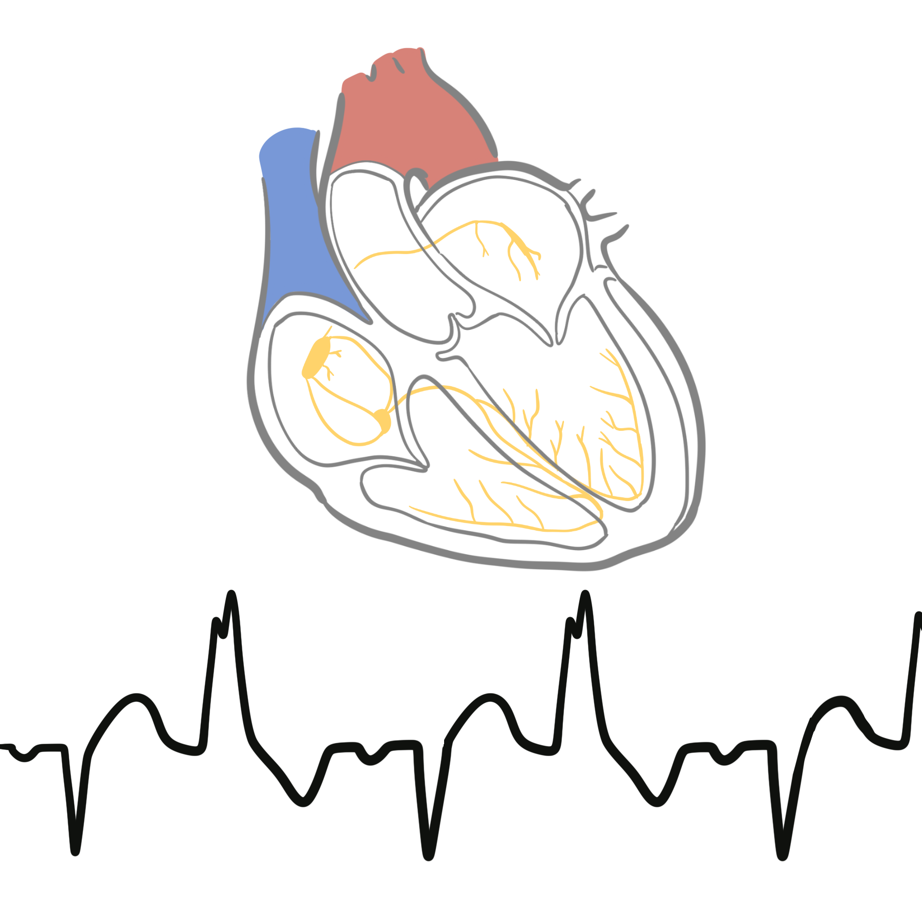
- Electrocardiographic abnormalities may include sinus tachycardia, atrial fibrillation, conduction abnormalities, non-specific ST–T changes or ventricular ectopy.
- Echocardiography commonly demonstrates left ventricular or biventricular dilatation, reduced ejection fraction, global hypokinesis, functional mitral regurgitation and elevated filling pressures.
- Intracardiac thrombus should be sought when ventricular dysfunction is severe.
- chest radiograph when congestion or another pulmonary process is suspected
- full blood count
- electrolytes, urea and creatinine
- liver function tests
- magnesium and phosphate
- blood glucose and glycated haemoglobin
- thyroid function
- iron studies
- B-type natriuretic peptide or N-terminal pro-B-type natriuretic peptide
- high-sensitivity troponin when acute myocardial injury is suspected.
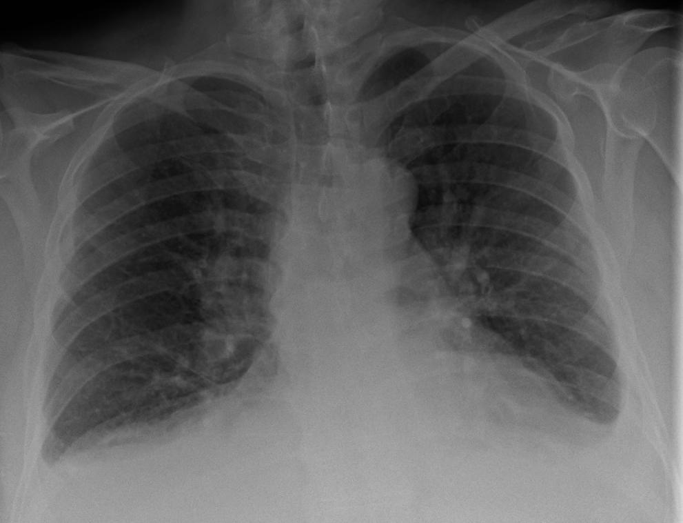
Chest X-ray showing cardiomegaly and pulmonary congestion. Image: James Heilman, MD, via Wikimedia Commons. Licensed under CC BY-SA 4.0.
What to notice
Detailed image description
Source: https://commons.wikimedia.org/wiki/File:CHF2016.png | Creator: James Heilman, MD | Licence: Creative Commons Attribution-ShareAlike 4.0 International (https://creativecommons.org/licenses/by-sa/4.0/) | Modifications: none.
Targeted Investigations
Depending on the presentation, consider:
- coronary computed tomography angiography or invasive coronary angiography to exclude clinically important coronary artery disease
- cardiac magnetic resonance imaging to define ventricular phenotype, assess fibrosis and evaluate myocarditis, infiltrative disease or another cardiomyopathy
- ambulatory electrocardiographic monitoring for palpitations, syncope or suspected arrhythmia
- genetic counselling and testing when there is a family history, conduction disease, ventricular arrhythmia or another feature suggesting inherited cardiomyopathy
- HIV, autoimmune, infectious or metabolic investigations when clinically indicated
- endomyocardial biopsy only when a specific diagnosis requiring histology is suspected and the result would alter management.2
A history of heavy alcohol consumption must not end the diagnostic evaluation. Coronary disease, genetic cardiomyopathy, tachyarrhythmia, hypertension and other myocardial toxins may coexist and may change management.1,2
Treatment
Alcohol Abstinence and Withdrawal Safety
Complete abstinence is the central disease-modifying intervention. Recovery of left ventricular function after abstinence is strongly associated with improved prognosis.1
Assess the risk of alcohol withdrawal before advising abrupt cessation. A patient with physiological dependence, previous withdrawal seizures or delirium tremens, severe comorbidity or limited support may require medically supervised withdrawal. Withdrawal treatment must be coordinated with addiction medicine or an appropriate inpatient service.4
Ongoing support may include:
- addiction-medicine review
- motivational and psychosocial interventions
- peer or family support
- nutritional assessment and thiamine replacement when indicated
- pharmacotherapy for alcohol-use disorder selected according to cardiac, hepatic and renal status
- relapse-prevention planning.
Heart-Failure Therapy
Treat heart failure with reduced ejection fraction according to current guideline-directed therapy. Unless contraindicated, foundational therapy includes:3
- Angiotensin receptor–neprilysin inhibitor, or an angiotensin-converting enzyme inhibitor or angiotensin receptor blocker
- Beta blocker
- Mineralocorticoid receptor antagonist
- Sodium–glucose cotransporter 2 inhibitor
Use loop diuretics to relieve pulmonary or peripheral congestion. Manage blood pressure, diabetes, iron deficiency, sleep-disordered breathing and other contributing conditions.
Medication initiation and titration should reflect blood pressure, renal function, potassium, volume status and clinical stability.
Arrhythmias and Device Therapy
Manage atrial fibrillation and other arrhythmias according to standard guidelines, including anticoagulation when indicated. Correct potassium and magnesium abnormalities and avoid additional cardiotoxic substances.
Decisions regarding an implantable cardioverter-defibrillator or cardiac resynchronisation therapy should follow standard heart-failure and arrhythmia criteria. When clinically safe, reassess ventricular function after abstinence and optimised medical therapy because reverse remodelling may alter device eligibility.1–3
Advanced Disease
Patients with persistent severe symptoms despite optimal therapy should be referred to a specialist heart-failure service for consideration of:
- advanced haemodynamic assessment
- temporary or durable mechanical circulatory support
- heart transplantation
- palliative and supportive care.
Sustained abstinence, treatment engagement and psychosocial stability are important parts of advanced-therapy assessment.
Improvement in ejection fraction after abstinence supports alcohol as an important causal contributor, but it does not justify stopping heart-failure therapy automatically. Persistent myocardial vulnerability and relapse risk require specialist reassessment.
Complications & Prognosis
Complications
- progressive heart failure
- acute pulmonary oedema
- cardiogenic shock
- functional mitral or tricuspid regurgitation
- atrial fibrillation
- ventricular tachycardia or ventricular fibrillation
- sudden cardiac death
- intracardiac thrombus and systemic embolism
- renal or hepatic dysfunction secondary to low output or congestion
- treatment-related hypotension, renal impairment or electrolyte disturbance
- alcohol withdrawal, seizures or delirium tremens during cessation.
Prognosis
Prognosis is strongly influenced by alcohol cessation and recovery of left ventricular function. Patients who abstain and develop meaningful reverse remodelling can have a substantially better outcome than those who continue heavy alcohol consumption.1
Poor prognostic features include:
- continued heavy alcohol use
- failure of left ventricular function to improve
- advanced heart-failure symptoms
- severe ventricular dilatation
- QRS prolongation or conduction disease
- atrial fibrillation or ventricular arrhythmia
- significant myocardial fibrosis
- renal, hepatic or nutritional complications
- recurrent non-adherence to treatment.
Repeat echocardiography is used to assess reverse remodelling after abstinence and guideline-directed therapy. Even when ejection fraction recovers, continued follow-up and sustained abstinence remain important because relapse or withdrawal of therapy may be followed by recurrent ventricular dysfunction.
References
- Domínguez F, Adler E, García-Pavía P. Alcoholic cardiomyopathy: an update. Eur Heart J. 2024;45(26):2294–2305. doi:10.1093/eurheartj/ehae362
- Arbelo E, Protonotarios A, Gimeno JR, Arbustini E, Barriales-Villa R, Basso C, et al. 2023 ESC Guidelines for the management of cardiomyopathies. Eur Heart J. 2023;44(37):3503–3626. doi:10.1093/eurheartj/ehad194
- McDonagh TA, Metra M, Adamo M, Gardner RS, Baumbach A, Böhm M, et al. 2021 ESC Guidelines for the diagnosis and treatment of acute and chronic heart failure. Eur Heart J. 2021;42(36):3599–3726. doi:10.1093/eurheartj/ehab368
- Haber PS, Riordan BC. Guidelines for the treatment of alcohol problems. 4th ed. Sydney: Specialty of Addiction Medicine, Faculty of Medicine and Health, University of Sydney; 2021. Available from: https://alcoholtreatmentguidelines.com.au


Members only discussions coming soon…