

Diverticular Disease

Overview
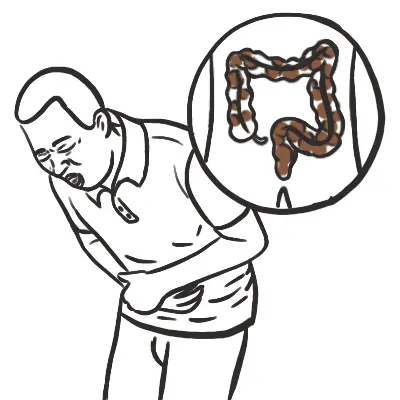
Diverticula of the colon consist of outpouchings of mucous membrane through the muscle wall of the bowel. In the colon, diverticula are found most commonly in the sigmoid and descending colon, and become increasingly rare in passing from the left to the right side of the colon. They are unusual before the age of 40 years, but they are found in about 30% of all autopsies in the elderly.
Definition
Diverticulum: abnormal sac-like protrusion from the wall of a hollow organ
Diverticulosis: presence of multiple diverticula, not symptomatic
Diverticulitis: inflammation of diverticula
Diverticular Disease: Complication of diverticulosis
True (congenital) diverticuli: contain all layers of colonic wall, often right-sided (i.e Merkel’s Diverticulum)
False (acquired) diverticuli: contain mucosa and submucosa, often left-sided (highest pressure)
Merkel’s Diverticulum: A true diverticulum, it is a reminant of proximal part of the yolk-stalk. May present with symptoms in a small majority of patients.
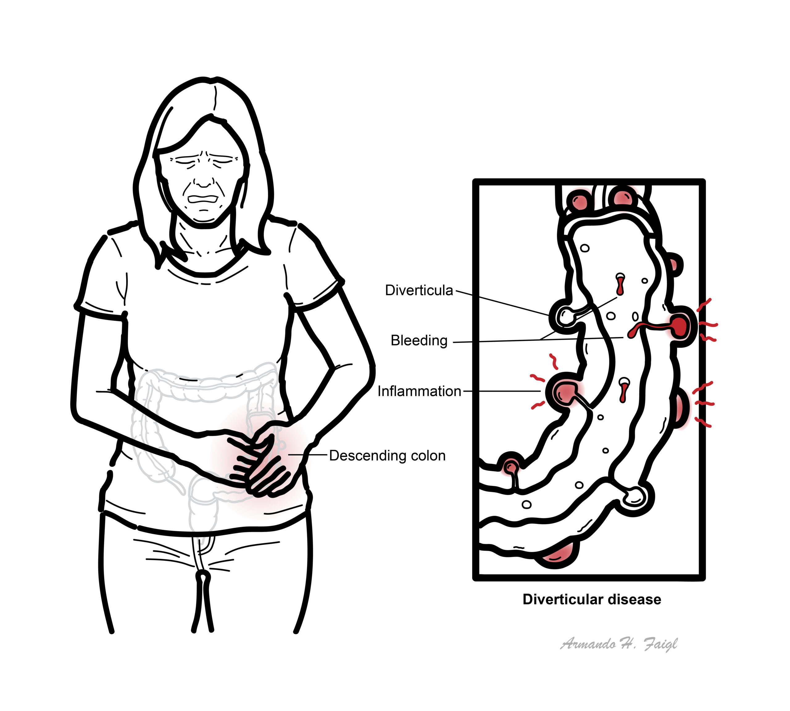
What to notice
Detailed image description
Colon Anatomy & Physiology
The colon is divided into:
- Caecum (appendix off this)
- Ascending colon
- Transverse colon
- Descending colon
- Sigmoid colon
- Rectum
The ascending and descending colon are retroperitoneal, these are immobile. The transverse is mobile and lies within the peritoneal cavity. The transverse colon is supported by the greater omentum superiorly and attaches to the posterior abdominal wall (in front of the retroperitoneal cavity) by the transverse mesocolon.
The transverse colon is attached to the greater curvature of the stomach and first part of the duodenum via the greater omentum. The sigmoid colon is also attached
Main features of large intestine structure:
- Complete layer of circular smooth muscle throughout, but incomplete bands of longitudinal muscle (taeniae coli) in colon (stops at the rectum)
- Fatty appendages along taeniae (appendices epiploicae)
- Folded internal mucosal appearances (haustrations)
- ‘Segmented’ external appearances (sacculations)
Blood supply – from superior and inferior mesenteric artery
- Superior mesenteric artery
- Ileocolic artery – last terminal ilealloop, caecum
- Right colic artery – ascending colon
- Middle colic artery – transverse colon up to the splenic flexure
- Inferior mesenteric artery
- Left colic – splenic flexure and descending colon.
- Sigmoid artery – sigmoid colon
- Superior rectal artery – rectum and upper anal canal
Autonomic nerve supply
- Sympathetic, mainly from greater splanchnic nerves via SMA and IMA plexuses.
- Parasympathetic, from vagus via SMA and IMA plexus from caecum to splenic flexure and from pelvic parasympathetics (S2, 3, 4) via hypogastric plexuses and retroperitoneal nerves from splenic flexure to upper anal canal
The colon is about 1.5 metres long, transverse being the longest and most mobile. The ascending and descending colon are fixed by the mesentery and do not move.
Risk Factors
| Risk Factors |
| Age >50 |
| Low fiber diet |
| Western diet |
| Obesity |
| NSAIDs |
Clinical Manifestations
Clinical Presentation of Diverticular Disease, one of four:
- Asympstomatic (majority)
- Painful Diverticular Disease
- Bleeding Diverticular Disease
- Diverticulitis.
Investigation
- FBC: leukocytosis, anaemia.
- CRP ± ESR.
- UEC/creatinine: dehydration and suitability for contrast imaging.
- LFTs and lipase where alternative diagnoses are possible.
- Urinalysis; pregnancy test where relevant.
- Blood cultures and lactate if febrile, hypotensive or septic.
Imaging
- CT abdomen and pelvis with IV contrast is the preferred test when:
- Diagnosis is uncertain.
- Symptoms are severe.
- The patient is systemically unwell.
- Complicated diverticulitis is suspected.
- The patient fails to improve.
- CT demonstrates bowel-wall inflammation and complications such as abscess, perforation, obstruction or fistula.
- Ultrasound or MRI may be considered when CT or IV contrast is unsuitable.
Colonoscopy
- Avoid colonoscopy during acute diverticulitis because of perforation risk.
- Consider colonoscopy after recovery, generally after 6–8 weeks, particularly following:
- Complicated diverticulitis.
- A first or atypical episode.
- Alarm symptoms such as bleeding, anaemia or weight loss.
On CT scan diverticulitis will have wall thickening (4mm) and surrounding oedema (inflammation) and signs of fat stranding.
Treatment
Management is mainly medical however there are indications of surgery (listed below).
Asymptomatic diverticulosis
- No specific medical treatment.
- Healthy balanced diet containing fibre, adequate fluid intake and regular exercise.
- Gradually increase fibre if constipation is present.
- Nuts, seeds, popcorn and fruit skins do not need to be avoided.
- Consider a bulk-forming laxative for constipation.
Symptomatic uncomplicated diverticular disease
- High-fibre diet or bulk-forming laxative.
- Paracetamol for pain.
- Consider an antispasmodic for abdominal cramping.
Acute uncomplicated diverticulitis
- Most clinically stable patients can be managed as outpatients.
- Oral fluids or a temporary light/liquid diet, advancing as symptoms improve.
- Paracetamol and adequate hydration.
- Selected immunocompetent, systemically well patients can be treated without antibiotics
Complicated diverticulitis
- Hospital admission.
- IV fluids, analgesia and IV antibiotics.
- Surgical or colorectal consultation.
- Treat sepsis and correct electrolyte abnormalities.
- Diverticular abscess:
- Smaller abscesses may respond to antibiotics.
- Consider image-guided percutaneous drainage for a larger accessible abscess—thresholds vary between guidelines, commonly around 3–5 cm.
| Indications for surgery |
| Failure of medical therapy |
| Wide-spread inflammation |
| Bowel obstruction |
| Abscess and fistula formation |
| Stenosis |
| Recurrent bleeding |
| Unacceptable symptoms for patients |
| Hinchey Staging and Treatment of Diverticulitis | ||
| Stage | Feature | Treatment |
| I | Phlegmon/small pericolic abscess (Localized) | Medical |
| II | Large abscess/fistula | Abscess drainage, resection +/- anastomsosis |
| III | Purulent peritonitis (ruptured abscess) | Hartmann procedure |
| IV | Feculent peritonitis | Hartmann procedure |
Complications & Prognosis
- Diverticular abscess or phlegmon — most common complication of acute diverticulitis.
- Perforation causing localised or generalised peritonitis.
- Sepsis and septic shock.
- Fistula formation (from perforation):
- Colovesical → pneumaturia, faecaluria or recurrent UTIs.
- Colovaginal → faecal vaginal discharge.
- Colocutaneous or entero-colonic fistula.
- Colonic stricture and bowel obstruction due to recurrent inflammation and fibrosis.
- Diverticular bleeding: often sudden, painless lower gastrointestinal bleeding; usually arises from diverticulosis rather than active diverticulitis.
- Recurrent episodes of acute diverticulitis.
- Persistent abdominal pain or altered bowel habit after inflammation resolves.
- Emergency surgery and temporary or permanent stoma may be required in severe perforation or uncontrolled sepsis.
- Most people with diverticulosis remain asymptomatic and never develop significant complications.
- Uncomplicated acute diverticulitis usually resolves with conservative treatment and has a generally good prognosis.
- Approximately 20% experience at least one recurrence within 10 years after an initial episode.
References
- National Institute for Health and Care Excellence. Diverticular disease: diagnosis and management. NICE guideline NG147. London: NICE; 2019.
- Qaseem A, Etxeandia-Ikobaltzeta I, Lin JS, Fitterman N, Shamliyan T, Wilt TJ, et al. Diagnosis and management of acute left-sided colonic diverticulitis: a clinical guideline from the American College of Physicians. Ann Intern Med. 2022;175(3):399–415. doi:10.7326/M21-2710.
- Peery AF, Shaukat A, Strate LL. AGA clinical practice update on medical management of colonic diverticulitis: expert review. Gastroenterology. 2021;160(3):906–911.e1. doi:10.1053/j.gastro.2020.09.059.
- Sartelli M, Weber DG, Kluger Y, Ansaloni L, Coccolini F, Abu-Zidan F, et al. 2020 update of the WSES guidelines for the management of acute colonic diverticulitis in the emergency setting. World J Emerg Surg. 2020;15:32. doi:10.1186/s13017-020-00313-4.




Members only discussions coming soon…