Overview
- An aneurysm is an abnormal localized dilatation of a blood vessel (>50% of its original diameter)
- It may be associated with structural abnormalities of collagen and elastin in the vessel wall
- Prevalence of abdominal aortic aneurysm: Much more common in females (9:1), 4% of men aged 65 (increasing with age).
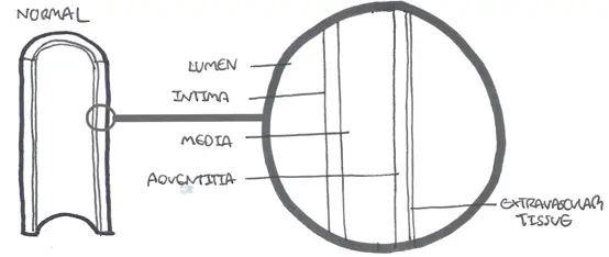
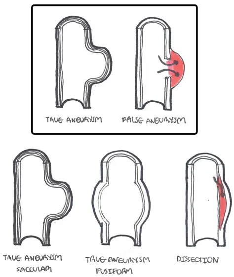
| Definition Aneurysm: an artery that has enlarged to greater than 1.5 times the expected diameter. True Aneurysm: The aneurysm is bound by all three layers of the vessel wall (intima, media and adventitia). The wall may be attenuated. False Aneurysm: Occurs when a blood vessel wall is injured, and the blood is contained by the surrounding tissues creating an apparent dilatation of vessel. Aortic dissection: occurs when a tear in the tunica intima of the aorta causes blood to flow between the layers of the wall of the aorta, forcing the layers apart. |
Types of Aneurysm
Aetiology
True Aneurysms
- Atheroma
- Infection (myocotic aneurysm in endocarditis, teritary syphilus -thoracic aneurysm)
- Connective tissue disorders (Marfan's Disease, Ethers-Donalos)
- Inflammatory (ie. Takuyasu Aortitis)
False Aneurysms
- Trauma
- Iatrogenic causes (ie. femoral cannulation, surgery)
Cerebral Aneurysm
A cerebral aneurysm is an acquired focal abnormal dilation of the wall of an artery in the brain. Intra-cranial aneurysms are most commonly located at branching points of the major arteries at the base of the brain, which course through the subarachnoid space. Cerebral aneurysms can lead to subarachnoid haemorrhage. saccular aneurysm of the middle cerebral artery is the most common
Thoracic Aortic Aneurysm
Often asymptomatic, diagnosed by widened mediastinum of chest x-ray or on CT/MRI. Rupture has high mortality and rare without prior symptoms
Abdominal Aortic Aneurysm
For more Information AAA
| Classic ruptured AAA triad: Hypotension/collapse, back/abdominal pain, Palpable/pulsatile abdominal mass. |
Often asymptomatic. ~40% are incidental findings (clinical examination, ultrasound, abdominal x-ray). Most common site is aneurysm below the renal arteries. 15% extend down to involve the origins of the common iliac arteries. AAA is associated with other peripheral aneurysm (popliteal). Risk of rupture and mortality increase with the diameter of the aneurysm.
| Remember AAA greater then 5.5cm in diameter need some for of intervention |
Management
Complications and Prognosis
- Rupture
- Thrombosis
- Embolism
- Fistulae
- Pressure on other structures
| Remember Classic triad of ruptured AAA congenital: Hypotension/collapse, back/abdominal pain, Palpable/pulsatile abdominal mass. |