Acute Lymphoblastic Leukaemia

Acute Lymphoblastic Leukaemia (ALL) is a malignancy of lymphoid cells. The two main ALL is T-cells ALL and B-cells ALL. In ALL there is uncontrolled proliferation of immature lymphoblasts in the bone marrow leading to overcrowding affecting other cell lineage. It is the commonest cancer in childhood, rare in adults and has a strong associated with Down’s Syndrome.
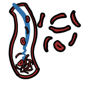
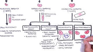
Leukaemia: Group of malignant disorder, affecting the blood forming tissue of the bone marrow lymph system and spleen
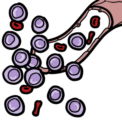
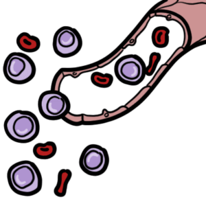
Acute lymphoblastic leukaemia: Most common leukaemia affecting children. Characterised by lymphoblasts in peripheral blood smear (lower cytoplasmic-nucleus ratio)
Acute Myeloid leukaemia: More common in young adults, adults. Characterised by the development of immature myeloblasts in the bone marrow.
Down’s Syndrome: Trisomy 21, a genetic disorder. Children with Down syndrome (DS) have an increased risk of B-cell precursor acute lymphoblastic leukemia.
| SUMMARY OF MAJOR LEUKAEMIA | ||
| Subtype | Description | Typical Group affects |
| Acute lymphoblastic leukaemia | Blast cells on peripheral blood smear or bone marrow aspirate | Children (53% <20yo) |
| Acute Myelogenous leukaemia | Blast cells on peripheral blood smear or bone marrow aspirate; Auer rods on peripheral smear, Myeloperoxidase positive | Adults |
| Chronic lymphoblastic leukaemia | Clonal expansion of at least 5,000 B lymphocytes per μL (5.0 × 10⁹ per L) in the peripheral blood | Older adults (85% >65yo) |
| Chronic Myelogenous leukaemia | Philadelphia chromosome (BCR-ABL1fusion gene) | Adults |
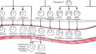
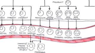
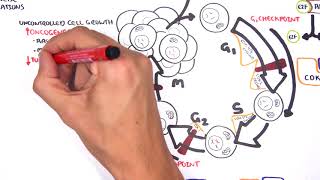
Haemtopoesis
ALL is classified based on 3 things:
Philadelphia chromosome is a balanced translocation between chromosome 9 and 22 which leads to the formation of the BCR/ABL fusion gene. This chromosome is seen mainly in Chronic myeloid leukaemia and sometime in ALL. In ALL it signifies a poorer prognosis.
| PROGNOSIS FEATURES IN CHILDHOOD ALL | ||
| Risk Factors | Favourable | Unfavourable |
| Age | 1-10yo | <1 or >10 yo |
| Gender | Female | Male |
| Race/ethnicity | Caucasian, Asian | Black, Hispanic |
| White blood cell count | <50000/ul | >50000/ul |
| Immunophenotype | B-precursor | T cell, Mature B cell |
| Genetic features | Hyperdiploidy, Translocation (12:21) | Philadelphia gene (9:22), Hypodiploidy |
| Extramedullary involvement | No | Yes |
Initial presentation
Bone marrow Failure
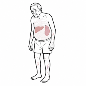
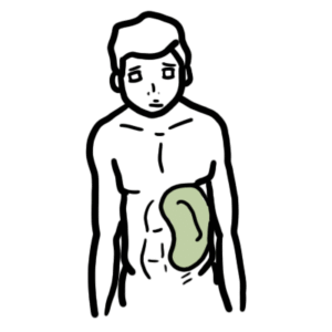
Infiltration (extramedullary haemtopoesis)
Chemotherapy Program
Haemotological remission means no presence of lymphoblasts on peripheral blood smear, normal bloods and <5% blasts in regenerating bone marrow.
Patient with cancer with recent chemotherapy (white cell count significantly down) spikes serious fever give antibiotic – Tazocin.
Prognosis
Acute Lymphoblastic Leukaemia and Acute Myeloid leukaemia
| DIFFERENCES BETWEEN ACUTE MYELOID LYMPHOBLASTIC LEUKAEMIA | ||
| Acute Myeloid Leukaemia | Acute Lymphoblastic Leukaemia | |
| Blast size on blood smear | Medium to large, uniform | Variable small to medium |
| Cytoplasm | Fine granules maybe present | Usualyl scant, a few coarse granules may be seen (morphology L3 – eosinophilic like) |
| Auer rods | Present | Absent |
| Nuclear chromatin | Finely Dispersed | Fine to corase |
| Genetic markers | Myeloid peroxidase present (only found in myeloid cells) | TDT marker present (only found in lymphoblasts) |


Discussion