

Aortic Aneurysm
Overview
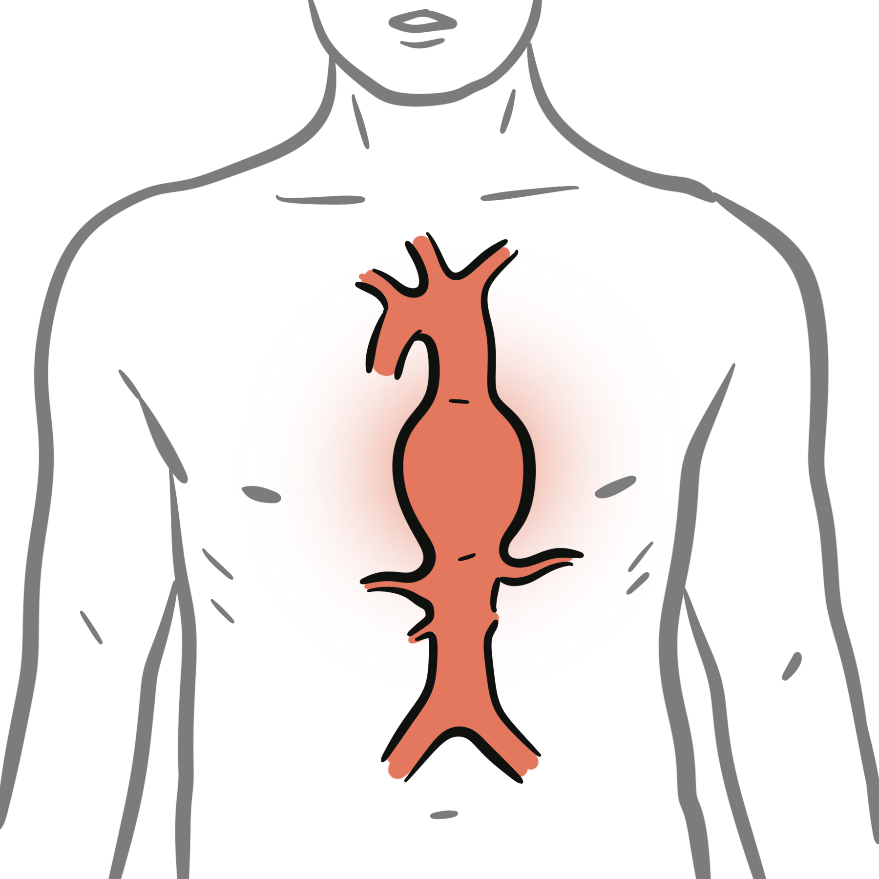
An aortic aneurysm is a pathological dilatation of the aorta caused by weakening of the arterial wall. It may involve the aortic root, ascending aorta, arch, descending thoracic aorta, thoracoabdominal aorta or abdominal aorta.
Most aortic aneurysms are asymptomatic and are discovered incidentally or through screening. Progressive enlargement increases the risk of rupture, dissection, thromboembolism and compression of adjacent structures. Risk is influenced by aneurysm location, diameter, growth rate, morphology, cause and individual patient characteristics.1–3
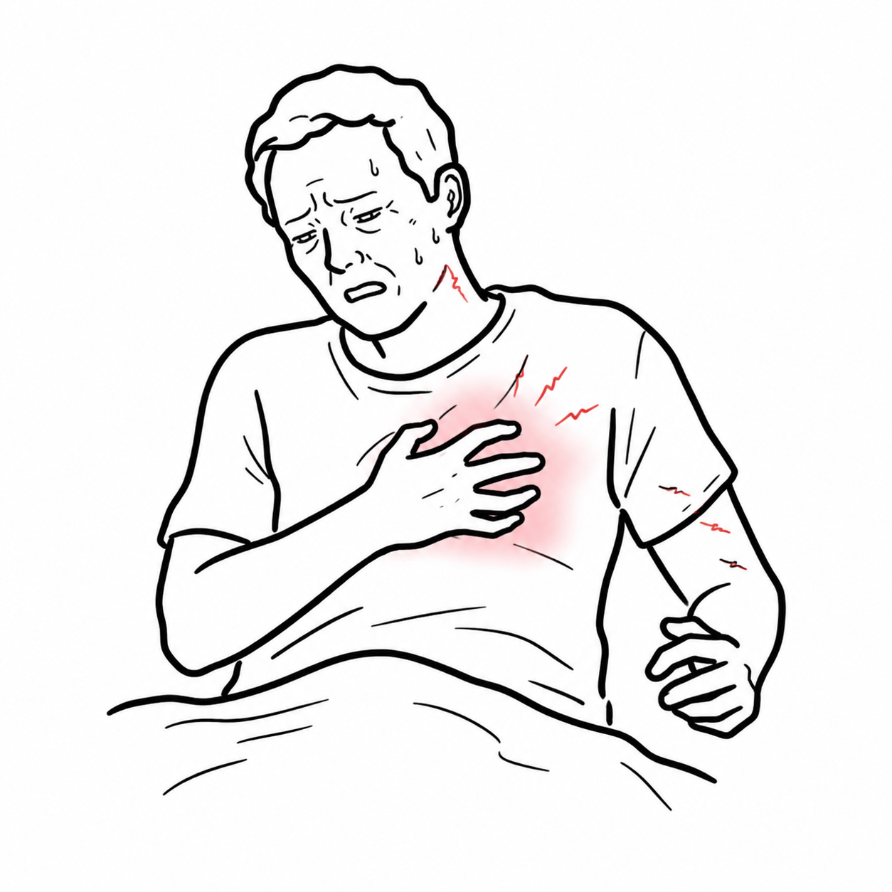
A new severe chest, back, abdominal or flank pain in a person with a known or suspected aneurysm must prompt urgent assessment for rupture or acute aortic syndrome.
Definition
- Aortic aneurysm
- Pathological dilatation of a segment of the aorta caused by loss of normal aortic-wall integrity.
- Thoracic aortic aneurysm
- An aneurysm involving the aortic root, ascending aorta, arch or descending thoracic aorta.
- Abdominal aortic aneurysm
- Dilatation of the abdominal aorta, most commonly involving the infrarenal segment; conventionally defined as a maximum diameter of at least 3.0 cm.
- Thoracoabdominal aortic aneurysm
- An aneurysm extending across the diaphragm and involving both the thoracic and abdominal aorta.
- Acute aortic syndrome
- A group of life-threatening aortic emergencies that includes aortic dissection, intramural haematoma and penetrating atherosclerotic ulcer.
Anatomy & Physiology
The aorta comprises:
- Aortic root.
- Ascending aorta.
- Aortic arch.
- Descending thoracic aorta.
- Thoracoabdominal aorta.
- Abdominal aorta.
The aortic wall has three layers:
- Intima: endothelial inner layer.
- Media: elastic fibres, collagen and smooth-muscle cells that provide strength and elasticity.
- Adventitia: connective tissue containing nerves and the vasa vasorum.
The aorta must accommodate pulsatile pressure while maintaining continuous blood flow. Its elastic properties permit expansion during systole and recoil during diastole. Wall stress increases as aortic radius and intraluminal pressure increase.
Aetiology & Risk Factors
Aetiology
Common causes and associations include:
- Degenerative and atherosclerotic aortic-wall disease, particularly in abdominal aneurysms.
- Age-related loss of elastin and medial structural integrity.
- Heritable thoracic aortic disease, including:
- Marfan syndrome.
- Loeys–Dietz syndrome.
- Vascular Ehlers–Danlos syndrome.
- Pathogenic variants involving genes such as ACTA2 and TGFBR1/2.
- Bicuspid aortic valve-associated aortopathy.
- Aortic coarctation.
- Previous aortic dissection.
- Inflammatory aortitis, including giant cell arteritis, Takayasu arteritis and IgG4-related disease.
- Infection of the aortic wall.
- Trauma or iatrogenic injury.
- Anastomotic pseudoaneurysm following previous vascular surgery.
Risk Factors
- Increasing age.
- Tobacco smoking, especially for abdominal aortic aneurysm.
- Male sex, although women with abdominal aneurysms may rupture at smaller diameters.
- Hypertension.
- Family history of aortic aneurysm or dissection.
- Established atherosclerotic cardiovascular disease.
- Hyperlipidaemia.
- Bicuspid aortic valve.
- Heritable connective-tissue or aortopathy syndrome.
- Previous aneurysm in another arterial territory.
- Rapid aortic enlargement.
- Chronic obstructive pulmonary disease.
Pathophysiology
Aortic aneurysm formation involves degradation of elastin and collagen, smooth-muscle cell loss, inflammation, oxidative stress and abnormal extracellular-matrix remodelling. These processes reduce the strength and elasticity of the aortic wall.
As the aorta enlarges, circumferential wall tension increases. This creates a cycle of progressive dilatation and further mechanical stress. Hypertension increases this stress, while smoking promotes inflammation and proteolytic degradation of the extracellular matrix.
Thoracic aneurysms often involve medial degeneration and may be associated with genetic aortopathy or bicuspid aortic valve. Abdominal aneurysms more commonly demonstrate transmural inflammation, atherosclerosis, proteolysis and intraluminal thrombus.
Aneurysm expansion may eventually cause:
- Rupture through the full thickness of the aortic wall.
- Intimal disruption and aortic dissection.
- Mural thrombus with distal embolisation.
- Branch-vessel obstruction or malperfusion.
- Compression of nearby structures.
Classification
Aortic aneurysms can be classified by location and morphology.
| Classification | Description |
|---|---|
| Aortic root or ascending aneurysm | Involves the root or ascending aorta; frequently associated with bicuspid valve or heritable aortopathy |
| Aortic arch aneurysm | Involves the transverse aortic arch |
| Descending thoracic aneurysm | Begins distal to the left subclavian artery and remains above the diaphragm |
| Thoracoabdominal aneurysm | Extends across the diaphragm and may involve visceral or renal arterial origins |
| Abdominal aortic aneurysm | Usually infrarenal but may be juxtarenal, pararenal or suprarenal |
| Fusiform aneurysm | Symmetrical dilatation involving the full circumference |
| Saccular aneurysm | Focal outpouching of part of the circumference; may carry greater concern for infection, ulceration or rupture |
| True aneurysm | Involves all three layers of the arterial wall |
| Pseudoaneurysm | Contained arterial-wall disruption in which blood is confined by surrounding tissues rather than all normal wall layers |
Clinical Manifestations
Most intact aortic aneurysms are asymptomatic.
Thoracic Aortic Aneurysm
Possible manifestations include:
- Chest or interscapular back pain.
- Aortic regurgitation from aortic-root dilatation.
- Heart failure symptoms.
- Hoarseness from recurrent laryngeal nerve compression.
- Cough, wheeze or dyspnoea from airway compression.
- Dysphagia from oesophageal compression.
- Facial or upper-limb swelling from venous compression.
- Neurological or upper-limb ischaemic symptoms from arch-vessel involvement.
- Features of an associated genetic syndrome or bicuspid aortic valve.
Abdominal Aortic Aneurysm
Possible manifestations include:
- A pulsatile abdominal mass.
- Persistent abdominal, flank, back or groin pain.
- Abdominal tenderness.
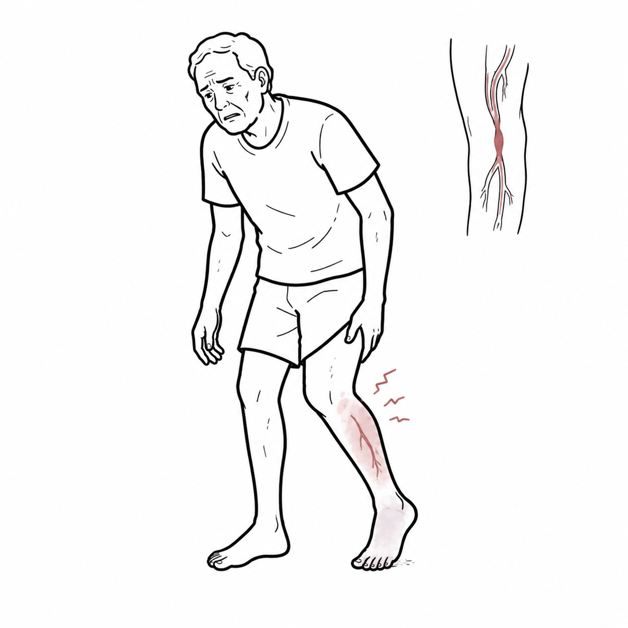
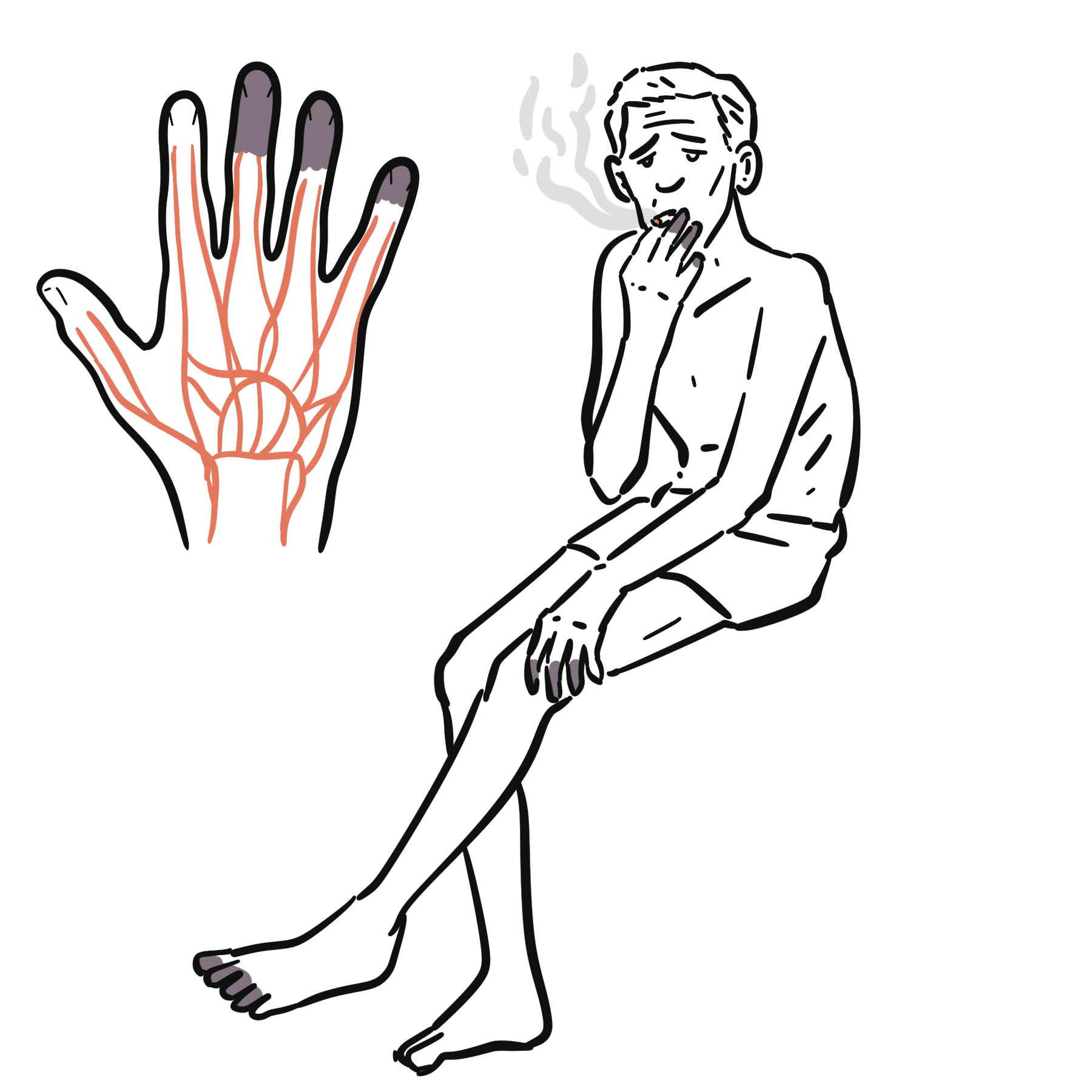
- Distal embolisation causing acute limb or digital ischaemia.
- Lower-limb ischaemia from thrombosis or associated occlusive disease.
- Ureteric, venous or gastrointestinal compression in large aneurysms.
Rupture or Acute Aortic Syndrome
Concerning features include:
- Abrupt, severe chest, back, abdominal or flank pain.
- Syncope or collapse.
- Hypotension or shock.
- A tender or expansile abdominal mass.
- New aortic regurgitation.
- Pulse or blood-pressure asymmetry.
- Focal neurological deficit.
- Acute limb, renal, mesenteric or spinal-cord ischaemia.
The classic triad of abdominal or back pain, hypotension and a pulsatile abdominal mass is important but is not present in every ruptured abdominal aortic aneurysm.
Aneurysm location helps explain the presentation. An ascending aneurysm may distort the aortic valve, an arch aneurysm may affect cerebral or upper-limb vessels, a descending thoracic aneurysm may compress the oesophagus or airway, and an abdominal aneurysm may embolise to the lower limbs.
Diagnosis & Investigations
Diagnosis requires demonstration of abnormal aortic dilatation on imaging. Assessment must establish:
- Aneurysm location and maximum diameter.
- Morphology and extent.
- Growth compared with previous imaging.
- Involvement of branch vessels.
- Presence of mural thrombus.
- Evidence of rupture, dissection, inflammation or infection.
- Associated aortic-valve disease.
- Suitability for open or endovascular repair.
Abdominal Ultrasound
Ultrasound is the preferred test for:
- Screening for abdominal aortic aneurysm.
- Confirming and measuring an uncomplicated infrarenal aneurysm.
- Serial surveillance when the aneurysm is adequately visualised.
It is inexpensive, non-invasive and does not involve radiation. It is less suitable for defining complex anatomy, branch-vessel involvement or suspected rupture.
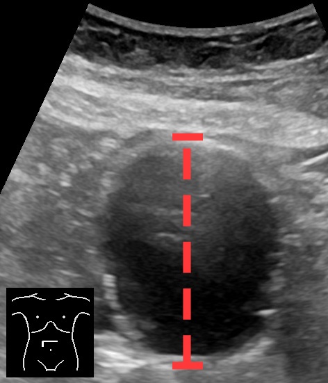
Axial ultrasound demonstrating an abdominal aortic aneurysm, with the anteroposterior diameter indicated. Image: Mikael Häggström, MD, via Wikimedia Commons. CC0 1.0.
What to notice
Detailed image description
Source: https://commons.wikimedia.org/wiki/File:Ultrasonography_of_abdominal_aortic_aneurysm_in_axial_plane,_annotated.jpg. Creator: Mikael Häggström, MD. Licence: CC0 1.0 Universal Public Domain Dedication. No modifications.
Echocardiography
Transthoracic echocardiography assesses:
- Aortic root and proximal ascending-aortic dimensions.
- Aortic-valve anatomy.
- Bicuspid aortic valve.
- Aortic regurgitation.
- Left-ventricular size and function.
It does not reliably visualise the entire thoracic aorta. Transoesophageal echocardiography can provide more detailed assessment in selected acute or perioperative settings.
CT Angiography
CT angiography provides detailed evaluation of:
- The entire aorta and branch vessels.
- Aneurysm morphology and maximum diameter.
- Calcification and mural thrombus.
- Rupture or retroperitoneal haemorrhage.
- Aortic dissection and malperfusion.
- Anatomy for endovascular or open repair.
Urgent CT angiography is generally appropriate when rupture or acute aortic syndrome is suspected and the patient is sufficiently stable for imaging. Imaging must not delay immediate surgical involvement or resuscitation.
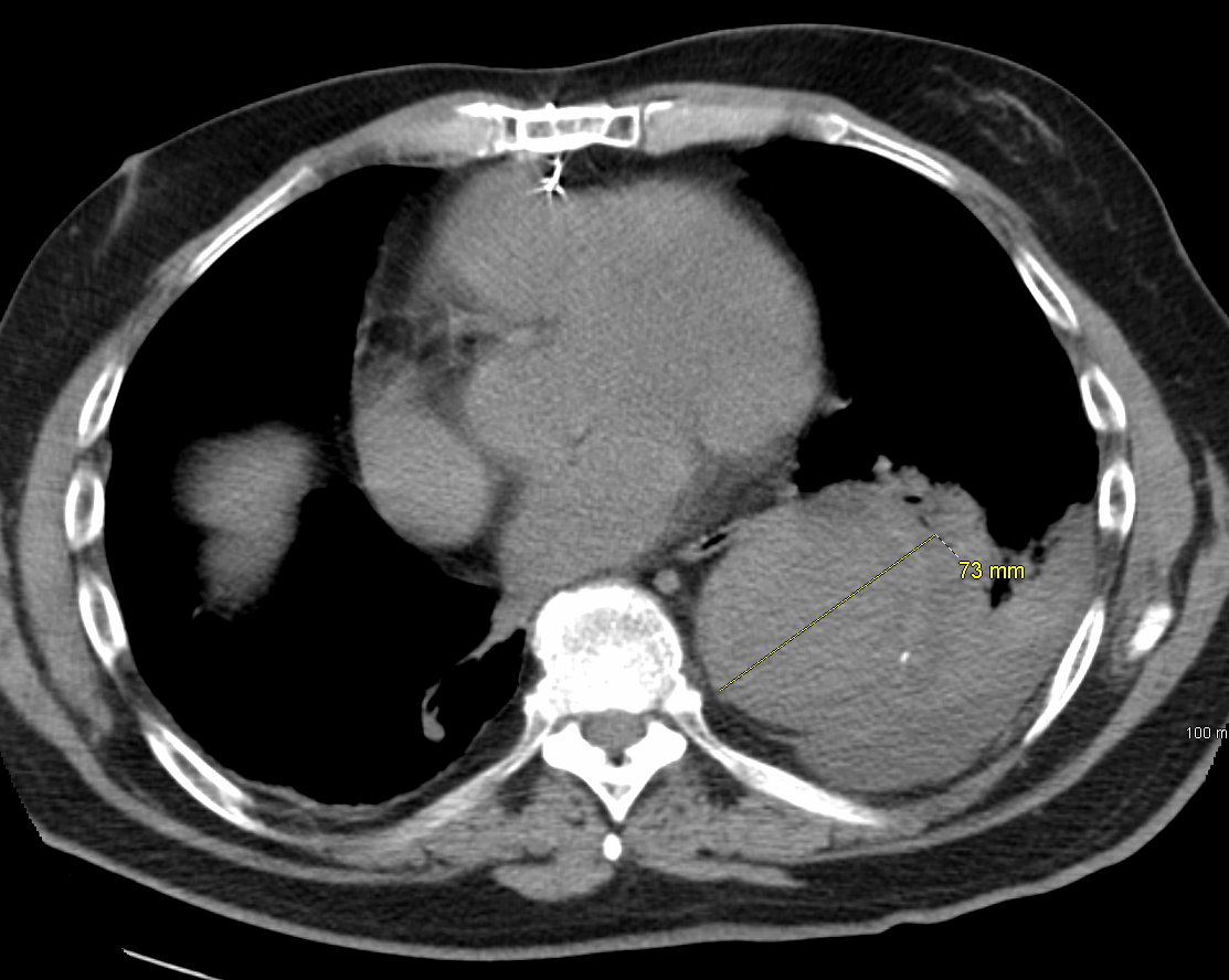
CT demonstrating a large thoracic aortic aneurysm measuring approximately 7 cm. Image: James Heilman, MD, via Wikimedia Commons. CC BY-SA 3.0.
What to notice
Detailed image description
Source: https://commons.wikimedia.org/wiki/File:CTAnurysmT.PNG. Creator: James Heilman, MD. Licence: Creative Commons Attribution-ShareAlike 3.0 Unported. No modifications.
Magnetic Resonance Angiography
MR angiography can provide cross-sectional imaging without ionising radiation and may be useful for:
- Younger patients requiring lifelong surveillance.
- Heritable thoracic aortic disease.
- Reducing cumulative radiation exposure.
- Selected patients who cannot receive iodinated contrast.
Measurement and Surveillance
Serial measurements should use a consistent anatomical landmark, imaging technique and measurement method wherever possible. Apparent growth may reflect differences in modality, cardiac phase, imaging plane or measurement convention.
For abdominal aortic aneurysm, guideline surveillance intervals commonly become shorter as diameter approaches the repair threshold. The ACC/AHA guideline recommends ultrasound:
- Every three years for an aneurysm measuring 3.0–3.9 cm.
- Annually for men with an aneurysm measuring 4.0–4.9 cm and women with an aneurysm measuring 4.0–4.4 cm.
- Every six months for men with an aneurysm measuring at least 5.0 cm and women with an aneurysm measuring at least 4.5 cm.1
For a stable sporadic thoracic aneurysm, repeat imaging is commonly performed after 6–12 months to establish growth rate and then every 6–24 months according to diameter, cause and proximity to an intervention threshold.1 Surveillance must be individualised for genetic aortopathy, bicuspid aortic valve, rapid growth and previous aortic intervention.
A normal abdominal ultrasound does not exclude thoracic aneurysm, aortic dissection or a contained aortic rupture. Choose imaging according to the suspected aortic segment and clinical urgency.
Treatment
Management depends on aneurysm location, diameter, growth, symptoms, morphology, cause, procedural risk and patient preference. Care should involve an experienced multidisciplinary aortic or vascular team.1–3
Suspected Rupture or Acute Aortic Syndrome
Immediate priorities include:
- Urgent vascular or cardiothoracic surgical involvement.
- Airway, breathing and circulation assessment.
- Large-bore intravenous access and cross-matched blood.
- Continuous haemodynamic monitoring.
- Appropriate analgesia.
- Rapid CT angiography when the patient is sufficiently stable.
- Avoidance of unnecessary delays or transfers.
- Immediate open or endovascular repair when indicated.
Resuscitation targets depend on the pathology. Uncontrolled hypertension worsens aortic-wall stress in acute aortic syndrome, whereas excessive crystalloid administration before haemorrhage control may worsen bleeding from a ruptured abdominal aneurysm.
Medical Management of an Intact Aneurysm
Medical management aims to reduce cardiovascular risk and aortic-wall stress:
- Complete smoking cessation.
- Appropriate blood-pressure control.
- Lipid-lowering therapy when atherosclerotic disease or another cardiovascular indication is present.
- Management of diabetes and other cardiovascular risk factors.
- Regular physical activity appropriate to the patient’s condition.
- Avoidance of extreme isometric exertion or heavy straining in patients with clinically important thoracic aortic dilatation.
- Adherence to scheduled surveillance imaging.
- Education about symptoms requiring emergency assessment.
Beta-blockers or angiotensin-receptor blockers may be used in selected thoracic aortopathies, particularly where hypertension or heritable disease is present. Treatment should be tailored to the underlying condition and current guideline recommendations.1
Elective Repair
Elective repair is considered when the risk of rupture or dissection exceeds the expected procedural risk.
Common indications include:
- Symptoms attributable to the aneurysm.
- Rapid enlargement.
- Saccular morphology or other high-risk anatomical features.
- Suspected infected aneurysm.
- Diameter reaching the recommended threshold.
- Lower thresholds in selected heritable aortopathies, bicuspid-valve disease or patients undergoing related cardiac surgery.
For uncomplicated infrarenal abdominal aortic aneurysm, repair is generally considered at approximately:
- 5.5 cm in men.
- 5.0 cm in women.
These thresholds are not absolute and must be interpreted alongside symptoms, growth, anatomy, operative risk and patient preference.1,2
For sporadic aortic-root or ascending-aortic aneurysm:
- Surgery is generally indicated at 5.5 cm.
- Repair may be reasonable from 5.0 cm in selected patients when performed by an experienced multidisciplinary aortic team.
- Earlier intervention may be indicated with rapid growth, symptoms, heritable aortopathy, bicuspid-valve risk factors or planned aortic-valve surgery.1
Rapid growth of a sporadic root or ascending aneurysm is defined by the ACC/AHA guideline as at least 0.5 cm in one year or at least 0.3 cm per year across two consecutive years. A threshold of at least 0.3 cm in one year applies to heritable thoracic aortic disease or bicuspid aortic valve.1
Repair Techniques
Options include:
- Open surgical repair: excision or exclusion of the aneurysmal segment with graft replacement.
- Endovascular aneurysm repair: placement of a stent graft through arterial access for suitable abdominal anatomy.
- Thoracic endovascular aortic repair: stent-graft treatment for suitable descending thoracic aneurysms.
- Complex endovascular repair: fenestrated or branched grafts for selected aneurysms involving visceral or renal branches.
- Hybrid repair: combined open and endovascular procedures in selected complex disease.
Endovascular repair usually offers lower early physiological stress but requires lifelong imaging surveillance for endoleak, graft migration, component separation and aneurysm-sac enlargement. Open repair has greater initial physiological impact but may provide greater long-term durability in suitable patients.
Complications & Prognosis
Complications
- Aneurysm rupture and haemorrhagic shock.
- Aortic dissection.
- Aortic regurgitation.
- Cardiac tamponade.
- Mural thrombosis and distal embolisation.
- Acute limb ischaemia.
- Renal, mesenteric, cerebral or spinal-cord malperfusion.
- Compression of adjacent structures.
- Aortoenteric or aortocaval fistula.
- Infection.
- Endoleak following endovascular repair.
- Graft migration, kinking, thrombosis or infection.
- Progressive dilatation of untreated aortic segments.
- Need for reintervention.
Prognosis
Poor prognostic features include rapid enlargement, symptoms attributable to the aneurysm, large diameter, saccular morphology, heritable aortopathy, uncontrolled hypertension, continued smoking and failure to attend surveillance.
Elective identification and planned repair have substantially better outcomes than emergency treatment after rupture. Even after successful repair, ongoing surveillance is required because residual aortic disease and procedure-related complications may develop.1–3
References
- Isselbacher EM, Preventza O, Hamilton Black J 3rd, Augoustides JGT, Beck AW, Bolen MA, et al. 2022 ACC/AHA guideline for the diagnosis and management of aortic disease: a report of the American Heart Association/American College of Cardiology Joint Committee on Clinical Practice Guidelines. Circulation. 2022;146(24):e334–e482. doi:10.1161/CIR.0000000000001106.
- Wanhainen A, Van Herzeele I, Bastos Goncalves F, Bellmunt Montoya S, Berard X, Boyle JR, et al. Editor’s choice—European Society for Vascular Surgery 2024 clinical practice guidelines on the management of abdominal aorto-iliac artery aneurysms. Eur J Vasc Endovasc Surg. 2024;67(2):192–331. doi:10.1016/j.ejvs.2023.11.002.
- Mazzolai L, Teixido-Tura G, Lanzi S, Boc V, Bossone E, Brodmann M, et al. 2024 ESC guidelines for the management of peripheral arterial and aortic diseases. Eur Heart J. 2024;45(36):3538–3700. doi:10.1093/eurheartj/ehae179.


Members only discussions coming soon…