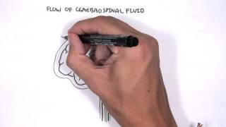
Hydrocephalus


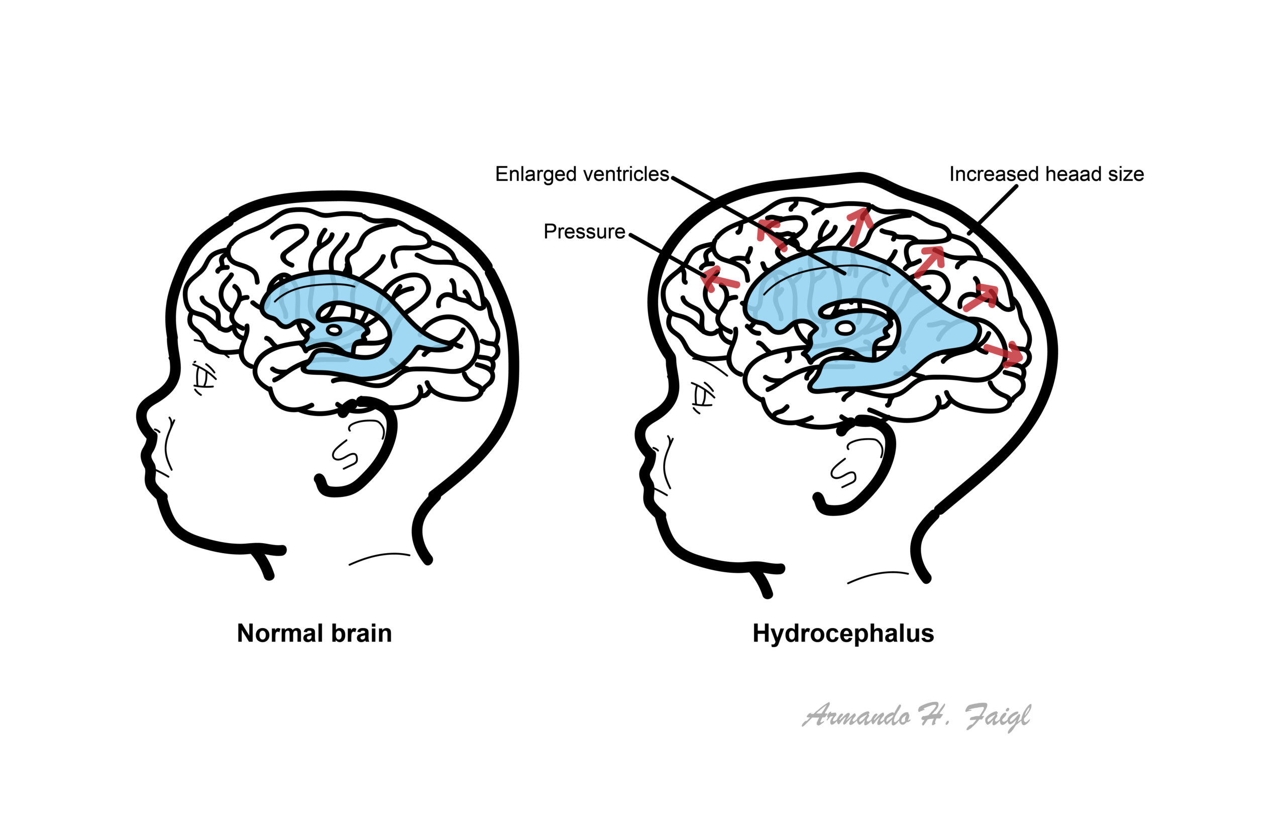
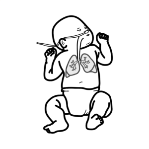
Hydrocephalus is excessive accumulation of CSF in the cerebral ventricles. In children, look for increasing head circumference, increased intracranial pressure, a bulging fontanelle, scalp vein engorgement, and paralysis of upward gaze. The most common causes include:
Treat the underlying cause, if possible; otherwise a surgical shunt is created to decompress the ventricles.
| Infantile Risk Factors |
| Birth weight <1500 g |
| Prematurity (gestational age ≤30 weeks) |
| Maternal diabetes |
| Low socioeconomic status |
| Male sex |
| Race/ethnicity (the risk is decreased in Asians) |
Excessive accumulation of cerebrospinal fluid in the brain that can be due to:
Many cases of hydrocephalus have both obstructive and absorptive components.
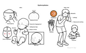
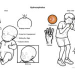
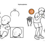
| DIFFERENT BETWEEN INFANT AND OLDER CHILDREN | |
| Infant | Older Child |
| Increasing head circumference, Macrocephaly | Headache (morning worse) |
| Delayed closure of the fontanel | Nausea and vomiting |
| Suture separation | Personality and mood changes |
| Bulging fontanel | Lethargy |
| Failure to thrive | Anorexia |
| Paresis of upward gaze (known as setting-sun sign) | Diplopia as a result of sixth-nerve palsy or third-nerve palsy with uncal herniation |
| Shrill cry | Papilledema |
Hydrocephalus should be suspected in an infant whose head circumference is enlarged at birth or in whom serial measurements cross percentiles in standard growth curves, indicating excessive head growth. Hydrocephalus should be considered in children with severe headache and other features suggesting increased intracranial pressure (ICP).
In a newborn, ultrasonography is the preferred technique for the initial examination because it is portable and avoids ionizing radiation. As the anterior fontanelle closes, the ultrasound is no longer a useful diagnostic modality.
In older infants and children with suspected hydrocephalus, CT or MRI should be performed. These imaging studies will also detect associated central nervous system (CNS) malformations or tumors.Brain imaging can help to distinguish obstructive (non-communicating) from absorptive (communicating) hydrocephalus.
Hydrocephalus can be divided up into congenital and acquired
Congenital Hydrocephalus are picked up on ultrasound
Acquired Hydrocephalus – usually occur later in life due to some sort of “event”
Hydrocephalus can be communicating or non-communicating
Hydrocephalus ex vacuo describes increases in CSF volume without increased CSF pressure, which is seen in conditions of reduced cerebral tissue (i.e malformation, atrophy).
Assume patients who present to emergency with headaches associated with a ventricular shunt, has infection/blockage. Refer as an emergency. Associated drowsiness is a particular pointer to blockage.
Shunt Complication
Royal Children’s Hospital Melbourne


Discussion