

Colorectal Cancer

Overview
Colon cancer is the second most commonly diagnosed cancer. 1/12 people will develop bowel cancer before the age of 85. However, there are Survival rates are increasing. Early bowel cancer is cured by surgery alone (screening is important!!). If untreated, or diagnosed when distance metastases are present, >98% pf patients die in <5 years.
Definition
Colorectal cancer: cancer arising from the colon, rectosigmoid junction or rectum.
Adenomatous polyp: premalignant gland-forming polyp that may progress to colorectal cancer.
Carcinoma in situ: abnormal malignant cells confined to the mucosa without invasion through the muscularis mucosae.
Mismatch-repair deficiency—dMMR: loss of normal DNA mismatch-repair function, producing microsatellite instability.
Total mesorectal excision—TME: surgical removal of the rectum together with its surrounding mesorectal tissue and lymphatic drainage.
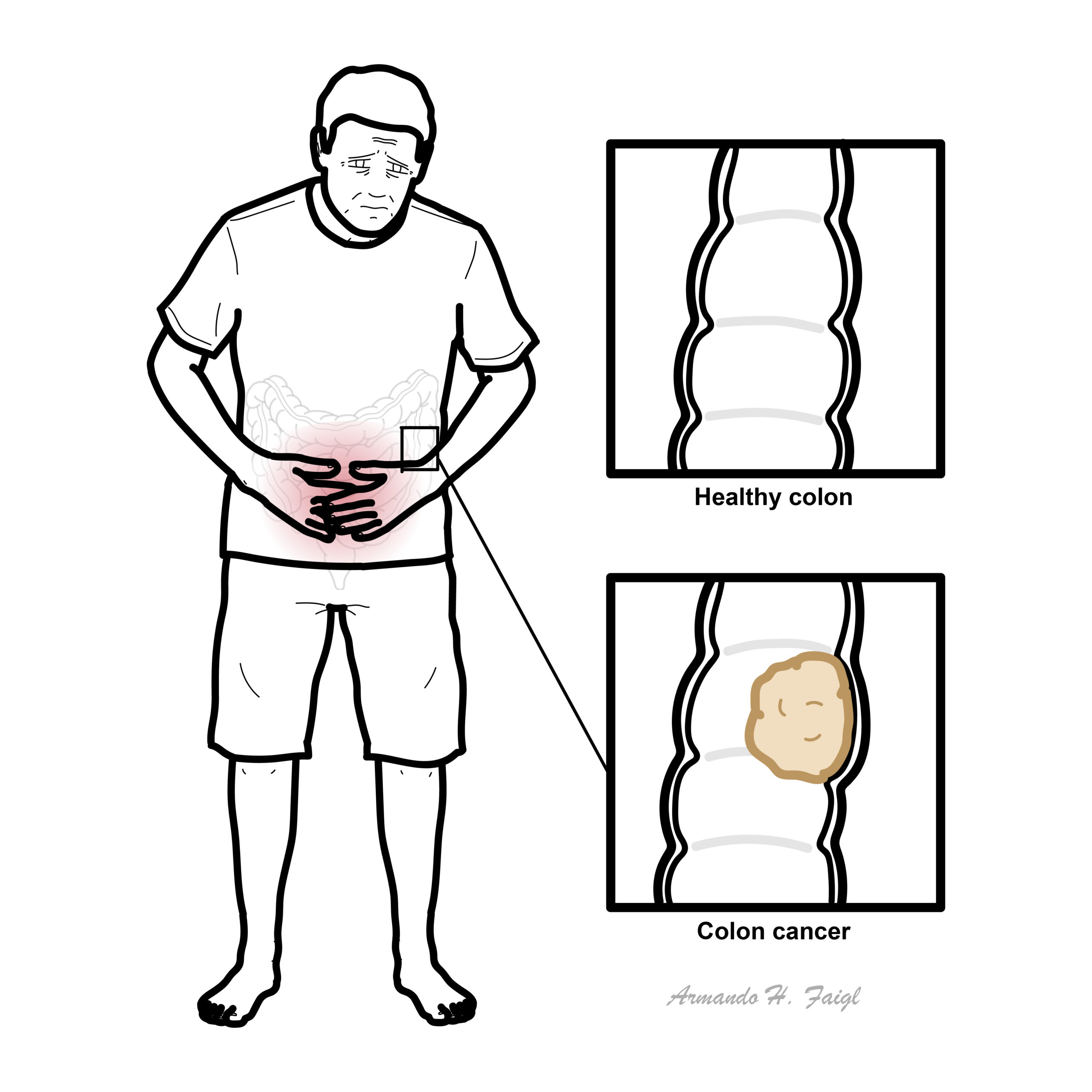
What to notice
Detailed image description
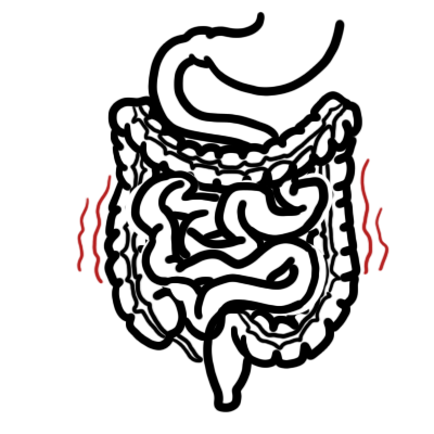
Colon Anatomy & Physiology
The colon is divided into:
- Caecum (appendix off this)
- Ascending colon
- Transverse colon
- Descending colon
- Sigmoid colon
- Rectum
The ascending and descending colon are retroperitoneal, these are immobile. The transverse is mobile and lies within the peritoneal cavity. The transverse colon is supported by the greater omentum superiorly and attaches to the posterior abdominal wall (in front of the retroperitoneal cavity) by the transverse mesocolon.
The transverse colon is attached to the greater curvature of the stomach and first part of the duodenum via the greater omentum. The sigmoid colon is also attached
Main features of large intestine structure:
- Complete layer of circular smooth muscle throughout, but incomplete bands of longitudinal muscle (taeniae coli) in colon (stops at the rectum)
- Fatty appendages along taeniae (appendices epiploicae)
- Folded internal mucosal appearances (haustrations)
- ‘Segmented’ external appearances (sacculations)
Blood supply – from superior and inferior mesenteric artery
- Superior mesenteric artery
- Ileocolic artery – last terminal ilealloop, caecum
- Right colic artery – ascending colon
- Middle colic artery – transverse colon up to the splenic flexure
- Inferior mesenteric artery
- Left colic – splenic flexure and descending colon.
- Sigmoid artery – sigmoid colon
- Superior rectal artery – rectum and upper anal canal
Autonomic nerve supply
- Sympathetic, mainly from greater splanchnic nerves via SMA and IMA plexuses.
- Parasympathetic, from vagus via SMA and IMA plexus from caecum to splenic flexure and from pelvic parasympathetics (S2, 3, 4) via hypogastric plexuses and retroperitoneal nerves from splenic flexure to upper anal canal
Aetiology & Risk Factors
Non-modifiable risk factors
- Increasing age
- Personal history of colorectal cancer or adenomatous polyps
- Family history of colorectal cancer
- Lynch syndrome
- Familial adenomatous polyposis—FAP
- Other hereditary polyposis syndromes
- Longstanding inflammatory bowel disease:
- Ulcerative colitis
- Crohn colitis
- Previous abdominal or pelvic radiotherapy
Modifiable risk factors
- High intake of processed meat
- High intake of red meat
- Low-fibre dietary pattern
- Obesity
- Physical inactivity
- Cigarette smoking
- Excessive alcohol consumption
- Type 2 diabetes
Colorectal cancer can still occur without an identifiable risk factor and should not be excluded solely because a patient is young.
Clinical Manifestation
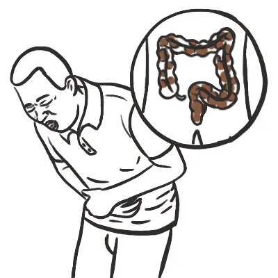
Signs and symptoms vary depending on the location of the tumour. 20% of people present at emergency with obstruction of large bowel or perforation.
| CLINICAL PRESENTATION | |||
| Right Colon | Left and Sigmoid Colon | Rectum | |
| Frequency | 20% | 75% | 5% |
| Pathology | Exophytic lesions wit occult bleeding | Annular, invasive lesions | Ulcerating |
| Symptoms | Weight los, weaknes, rarely obstruction | Constipation, change in bowel habits, abdominal pain, rectal bleeding | Obstruction, tenesumus, rectal bleeding |
| Signs | Fe+ deficiency anaemia, QLR mass (10%) | Palpable mass on Digital Rectal Examination | |
Elderly persons who present with iron-deficiency anemia should be investigated for colon cancer.
Occult blood in the stool of a person older than 40 years should be considered colon cancer until proven otherwise. To rule out colon cancer, perform a colonoscopy.

Clinical Examination
- Anaemia
- Palpable mass on abdominal examination
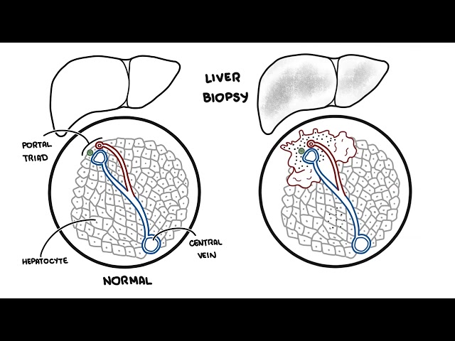
- Palpable nodular liver (metastasis)
- Pigmentation (Peutz-Jeghers syndrome)
- Discrete black brown lesion on lips
Metastatic disease clinical manifestation
- Liver metastases:
- Right upper-quadrant pain
- Hepatomegaly
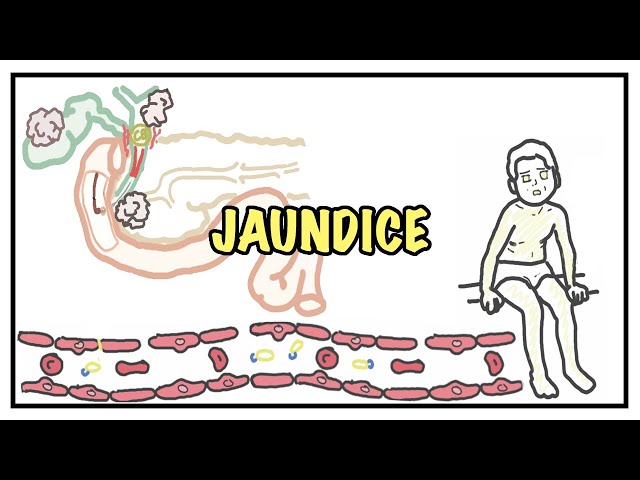
- Jaundice
- Lung metastases:
- Cough
- Dyspnoea
- Peritoneal metastases:
- Ascites
- Bowel obstruction
- Bone or neurological symptoms are less common.
For more information: Clinical Presentation of Per-rectal bleeding
Diagnosis & Investigations
- FBC: iron-deficiency anaemia
- Iron studies
- UEC and renal function
- LFTs
- Coagulation studies
- Carcinoembryonic antigen—CEA:
- Colonoscopy
- Principal diagnostic investigation.
- Allows biopsy of the tumour.
Diagnosis: Histopathology
- Colorectal cancer are primarily adenocarcinoma.
- Colon cancer can be polyploid, ulcerative, stenosing or infiltrative.
- All newly diagnosed colorectal cancers should generally undergo MMR protein immunohistochemistry or MSI testing.
- Abnormal results may:
- Suggest Lynch syndrome
- Influence prognosis
- Affect adjuvant-treatment decisions
- Predict response to immunotherapy
- Metastatic disease requires molecular testing, commonly including:
- KRAS and NRAS
- BRAF V600E
- MSI/MMR status
- HER2 in selected RAS/BRAF wild-type cancers
- Other actionable alterations where comprehensive profiling is available.
- Abnormal results may:
Screening for colon cancer is available: FOBT. Done yearly after 50yo (Australia).
Pathophysiology
Staging
- CT chest, abdomen and pelvis
- Complete colonoscopy
- Baseline CEA
- MRI liver if liver lesions remain uncertain or potentially resectable
TNM staging
- T: depth of invasion through the bowel wall and adjacent structures
- N: regional lymph-node involvement
- M: distant metastatic disease
| Stage | General description |
|---|---|
| 0 | Carcinoma in situ, confined to the mucosa |
| I | Invades the submucosa or muscularis propria; no nodal disease |
| II | Extends through the bowel wall; no regional lymph-node metastases |
| III | Regional lymph-node involvement |
| IV | Distant metastatic disease |
Treatment
Treatment depends on tumour location, stage, molecular profile, operability, performance status and patient preference. Management should occur through a colorectal multidisciplinary team.
Stage 0
- Endoscopic polypectomy or local excision if completely removable.
- Segmental colectomy if the lesion cannot be adequately removed endoscopically.
Stage I
- Surgical resection of the affected colon segment with regional lymph-node removal.
- Adjuvant chemotherapy is not routinely required.
- Cure rates are generally high.
Stage II
- Surgical resection is the principal treatment.
- +/-chemotherapy
Stage III
- Surgical resection followed by adjuvant chemotherapy.
- Common regimens:
- FOLFOX: fluorouracil, leucovorin and oxaliplatin
- CAPOX: capecitabine and oxaliplatin
Rectal cancer treatment
- Early rectal cancer
- Selected small, favourable T1 tumours may undergo transanal local excision.
- Most stage I cancers require oncological rectal resection with total mesorectal excision.
- Surgical options include:
- Low anterior resection
- Coloanal anastomosis
- Abdominoperineal resection when sphincter preservation is not possible.
- Locally advanced rectal cancer
- Total neoadjuvant therapy: provides systemic chemotherapy and pelvic radiotherapy before surgery.
- Surgery
Metastatic colorectal cancer
- Chemotherapy
- FOLFOX
- CAPOX
- FOLFIRI
- FOLFOXIRI in selected fit patients
- Targeted therapy
- Anti-VEGF therapy, such as bevacizumab
- Anti-EGFR therapy, such as cetuximab or panitumumab:
- Only for RAS wild-type tumours
- Most effective in selected left-sided primary cancers
- BRAF-targeted therapy for BRAF V600E disease
- HER2-targeted therapy for selected HER2-positive disease
- KRAS G12C-directed therapy for selected previously treated disease
- Immunotherapy
- Palliative treatment
- Radiotherapy for pain, bleeding or local obstruction
- Colonic stent or diverting stoma for obstruction
Complications & Prognosis
Disease-related complications
- Large-bowel obstruction
- Bowel perforation
- Venous thromboembolism
- Metastasis
Treatment-related complications
- Surgery
- Anastomotic leak
- Intra-abdominal infection
- Ileus or bowel obstruction
- Stoma complications
- Incisional hernia
- Chemotherapy
- Myelosuppression and infection
- Nausea, diarrhoea and mucositis
- Oxaliplatin-associated peripheral neuropathy
- Hand–foot syndrome from capecitabine
- Cardiotoxicity from fluoropyrimidines
- Pelvic radiotherapy
- Diarrhoea and proctitis
- Urinary dysfunction
- Sexual dysfunction
- Infertility
Prognosis
- Early-stage colorectal cancer is frequently curable with surgery.
- Regional lymph-node disease carries a greater recurrence risk but can still be cured with surgery and adjuvant therapy.
- Metastatic disease is usually not curable, although selected patients with resectable liver or lung metastases may achieve long-term survival.
- Australian five-year relative survival across all colorectal-cancer stages is approximately 72%; stage-specific outcomes are substantially better for localised disease and poorer for metastatic disease.
- Duke’s staging looks at the 5 year survival rate and this is based on the layers/sites the tumours have invaded.
- Duke’s staging (5 year survival rate)
- Stage A – Tumour is confined to bowel wall only (75-90%)
- Stage B – Tumour is through bowel wall (55-70%)
- Stage C – Tumour has spread through regional lymph nodes and has a 30% 5 year survival rate
- Stage D is where the tumour has metastasized has a 5% 5 year survival rate.
- Duke’s staging (5 year survival rate)
Screening & Prevention
- Australia’s National Bowel Cancer Screening Program provides faecal immunochemical testing for eligible people aged 45–74 years.
- A positive screening test requires colonoscopy; it does not itself diagnose cancer.
- People with symptoms, inflammatory bowel disease, strong family history or hereditary syndromes require individualised assessment rather than relying on routine population screening.
- Risk reduction includes:
- Maintaining a healthy weight
- Regular physical activity
- Avoiding smoking
- Limiting alcohol and processed meat
- Consuming adequate dietary fibre.
Family Adenomatous Polyposis
Overview
- Autosomal dominant
- APC gene on chromosome 5q21
- Penetrance 100%
- Characterized by 100’s of colonic polyps
- Mean age 39
Other manifestations
- Periemullary polyps
- Congenital hypertrophy of retinal pigment epithelium
- Osteomas skill and mandible
- Dental abnormalities
Hereditary Non-Poyposis colorectal Cancer
Overview
- Also known as Lynch Syndrome
- Autosomal dominant
- 80% lifetime risk of developing colorectal cancer
- increased risk uterine, gastric, urinary tract, small bowel and brain cancer
- Defect in DNA mismatch repair cause microsatellite instability
Peutz-Jeghers Syndrome
Overview
- Characterized by the development of noncancerous growths called hamartomatous polyps in the gastrointestinal tract (particularly the stomach and intestines)
- Greatly increased risk of developing certain types of cancer
- Increases risk of small-bowel and pancreatic cancer, colorectal cancer and sex-cord tumours with annular tubules of the ovary
- Other clinical features often found in children and young adults: Perioral pigmentation, pigmentations of fingers
References
- Cancer Council Australia. Clinical practice guidelines for the prevention, early detection and management of colorectal cancer [Internet]. Sydney: Cancer Council Australia; 2026.
- PDQ Adult Treatment Editorial Board. Colon cancer treatment (PDQ): health professional version [Internet]. Bethesda: National Cancer Institute; updated 2025 Feb 12.
- PDQ Adult Treatment Editorial Board. Rectal cancer treatment (PDQ): health professional version [Internet]. Bethesda: National Cancer Institute; updated 2025 Feb 12.
- Cervantes A, Adam R, Roselló S, Arnold D, Normanno N, Taïeb J, et al. Metastatic colorectal cancer: ESMO Clinical Practice Guideline for diagnosis, treatment and follow-up. Ann Oncol. 2023;34(1):10–32. doi:10.1016/j.annonc.2022.10.003.
- Cancer Australia. Bowel cancer—colorectal cancer—in Australia statistics [Internet]. Canberra: Australian Government; updated 2025 Dec 18.





Members only discussions coming soon…